Non-invasive determination of differentiation / undifferentiation for culturing while maintaining undifferentiated hPSC cells
Point
Non-invasive phase contrast imaging and analysis allows for hPSC (such as hiPSC and hESC) colonies to be distinguished on the basis of differentiation state.
Overview
A large quantity of undifferentiated hPS cells are required when inducing differentiation into a given target cell type. Furthermore, culture conditions and other factors may induce unintended differentiation, and differentiated cells will need to be removed to prevent mixing with undifferentiated colonies. The only methods for identifying the occurrence of differentiation are staining with biological markers for the differentiated state, and morphological characterization. Staining is an invasive inspection method that is especially problematic because stained cells cannot usually continue in culture. Morphological characterization may also be problematic due to the use of subjective criteria, and dependence on operator judgment.
Current
Issue-1
Staining method is invasive
Differentiated colonies can often be accurately distinguished when staining for biological markers of differentiation and/or undifferentiation, but this is not a completely reliable method.
Due to the invasiveness of cell staining, it is difficult to continue culturing stained cells following evaluation. Additionally, staining reagents are expensive.
Issue-2
Experience is required to accurately judge colony shape.
Distinguishing differentiated cells on the basis of colony morphology is non-invasive, but judgment varies among operators since it's informed by visual observation. In addition, extensive culture experience is required to make accurate judgments.
Solution
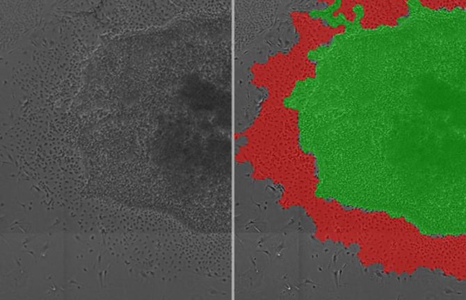
If a non-invasive method is used to identify hPSC differentiation state, then undifferentiated cells can continue to be used for culture.Analysis of phase contrast images of hPSC colonies allows for differentiated and undifferentiated areas within colonies to be distinguished on the basis of cell size and texture. Uniform criteria can be used for analysis of a wide range of images. It is also possible to quantify the number of differentiated colonies in order to compare the impact of different culture conditions and other factors.
Using the BioStudio-T*, a cell observation system that can be installed inside an incubator, non-invasive phase contrast images can be captured over an extended period without perturbing the culture environment.
*This product has been discontinued. If you have any questions about cell cultures, assays, or products, please contact us using the contact form.Cell image analysis software Cell Analysis Module
Cell image analysis software
NIS-Elements Cell Analysis Module
The following application softwares are for the purpose that specific image analysis can be performed simply using Nikon's Image Analysis Software in the field of research and development. As we have developed image analysis algorithms for many years and accumulated know-how, we have started to provide add-on Modules.
Click here for detailsNikon will contribute to solving your cell culture issues with its image analysis
techniques and know-how on cell quality evaluation.
Click hereInquiry Form
Share this page