
MPS observation
MPS observation
MPS observation
MPS observation
The world revealed by “imaging with cell assays”
MPS (microphysiological systems) such as OoC (organ-on-a-chip) are systems that allow a physiologically reproducible assay by mimicking the complexity of the three-dimensional structures and functions of living organisms in vitro. By observing the inside of an MPS, it is thought that phenomena that occur inside the human body can be better understood.
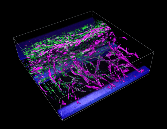
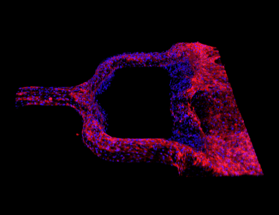
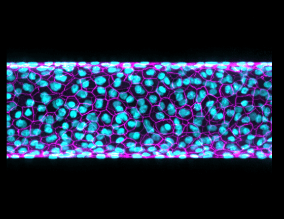
Time-lapse imaging analysis of angiogenesis induction using a 3D angiogenesis model
Sample: 3D angiogenesis structure (in vitro perfusion angiogenesis model) using MIMETAS’s “OrganoPlate® 3-lane 40”, the 3D tissue culture platform
Imaging product: AX/AX R confocal microscope, 40X water immersion objective (NA 1.25)
Description of colors: Green: Actin, Red: CD31 (vascular endothelial marker), Blue: DAPI (nuclei)
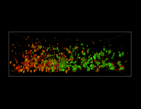
Confocal imaging of CAR-T cell dynamics
Sample: Simple 3D immune cell-mediated killing assay model using AIM Biotech’s 3D cell culture chip built to measure the immune effects of T cells by in vitro imaging
Imaging product: AX/AX R confocal microscope, 20X water immersion objective (NA 0.95)
Description of colors: Green: GFP labeled Hep3B, Orange: mCherry labeled CD133 specific CAR-T cells, Red: Alexa Fluor 633 labeled cleaved Caspase-3

Analyze the effects of drugs in vivo using cell cultures in fine fluid channels and structures.

Expected to be an alternative to conventional animal testing.

Support MPS observation by our microscopes and software suitable for image acquisition and analysis of complex 3D structures that imitate living organisms.
Application Notes
Application notes for MPS observation.
 Confocal Imaging of CAR-T Cell Dynamics Using an Organ-on-a-chip Platform
 Time-lapse imaging analysis of angiogenesis induction using a 3D model
 Selecting the Right Objectives - Bright, Sharp Imaging of Structures down to Deep Areas
 Capturing the physiological complexity of human tissues in vitro with organ-on-a-chip
 3D Imaging of Intestinal Organoid
 Quantitative 3D Imaging of Living Organs-on-Chips with a High-Speed Point-Scanning Confocal System
 Live Imaging of Paneth Cell Secretory Responses in Innate Immunity by Using Three-Dimensional Culture of Small Intestinal Epithelial Cells
Glossary
Terms related to imaging of MPS, organoids, and OoC, and also includes observation examples relating to each term.
Nikon BioImaging Labs
The Nikon BioImaging Lab operates in three countries around the world as an open facility supporting solutions to drug discovery research issues.
Nikon BioImaging LabsSolutions
Introduces a method for quantitative analysis of 3D cell culture systems such as spheroids, organoids, and organs-on-chips at high speeds and with deep imaging.
Related SolutionsRelated Products
Introduces hardware and software suitable for MPS observation