Diabetic retinopathy is the major cause of vision loss worldwide. Nikon Ophthalmology Solutions and Verily Life Sciences LLC* (United States; referred to below as "Verily", formerly known as Google Life Sciences) with Google Inc. as its parent company, are collaborating on the development of a of diagnosis support system using artificial intelligence (AI) for early diagnosis. Here, we introduce our challenge.
- *Verily Life Sciences LLC: Verily is focused on using technology to better understand health, as well as prevent, detect, and manage disease. Our mission is to bring together technology and life sciences to uncover new truths about health and disease.
Diabetic retinopathy: early diagnosis is important
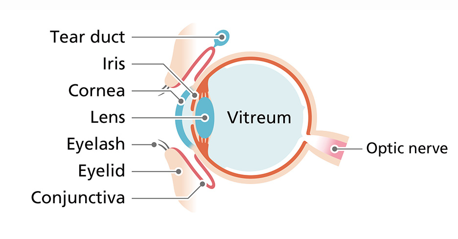
Diabetic retinopathy which is a complication of diabetes, is one of the reasons many people lose their vision. The retina consists of thin-film tissues of the ocular fundus*, sensing light and colors and delivering the information to the brain. Weakening vision due to retina damage will eventually lead to vision loss, known as diabetic retinopathy. However, early diagnosis and appropriate treatment can reduce the disease's progress.
- *Ocular fundus: Tissues at the base of the eyeball.
Finding initial-stage symptoms at an early stage by effectively using ultra-widefield retinal images
Diabetic retinopathy is a disease that is difficult to detect in its initial stages. But if there is a solution to support doctors who read*1 retinal images, there are possibilities to realize early diagnosis.
In a joint project with Verily Life Science LLC, we collect retinal images taken with an ultra-widefield retinal imaging device*2 from medical facilities all over the world. Multiple experts read the progress of disease according to strict standards. We employ AI to learn from this diagnostic data from medical experts and the accumulated retinal images, which supports diagnosis of diabetic retinopathy.
- *1Image reading: Examination using images
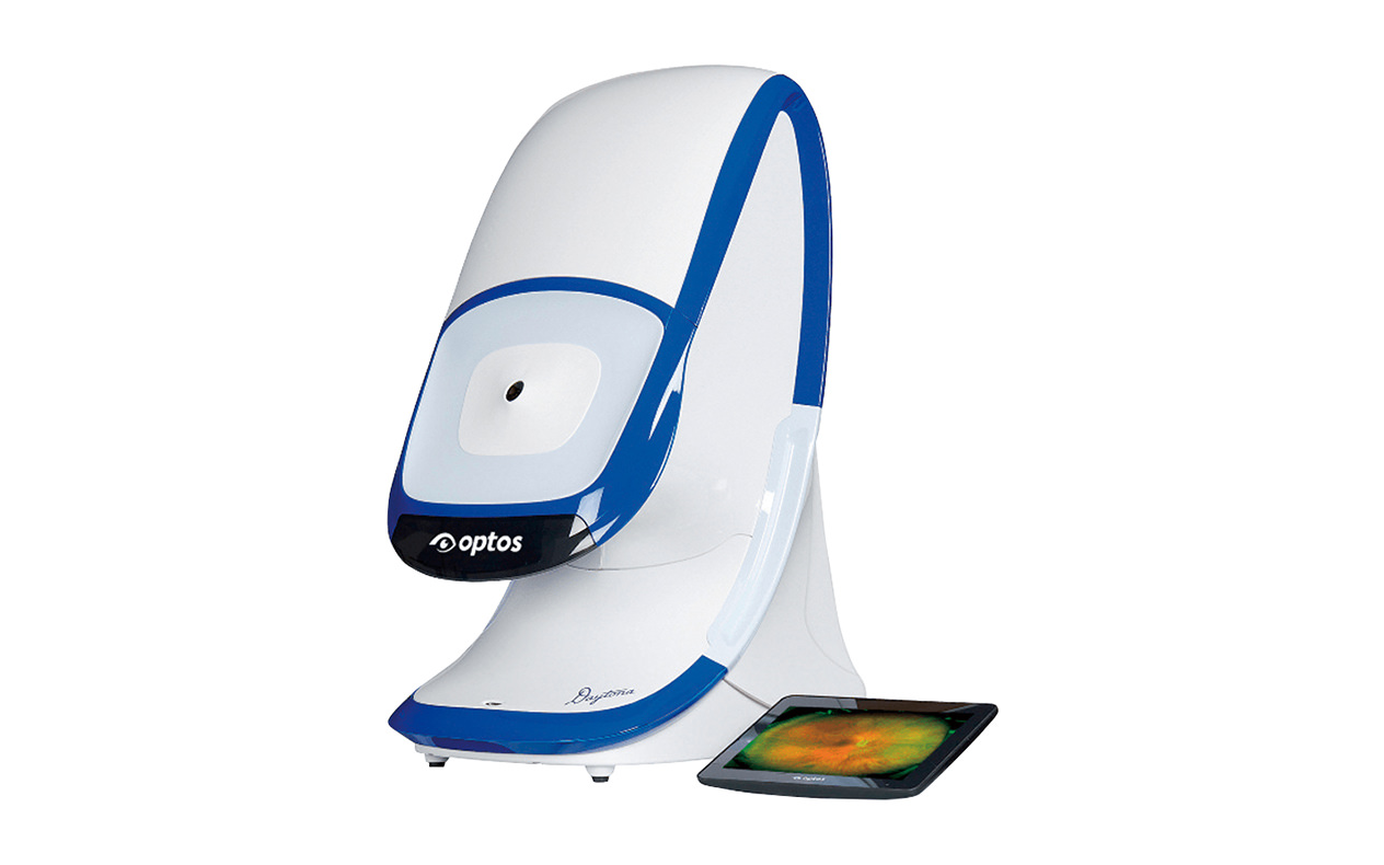
- *2ultra-widefield retinal imaging device: A product from the Nikon Group's Optos. It captures a digital image of the retinal area with its wide field of view of 200˚ simultaneously.
Aiming for early diagnosis of various diseases
Our ongoing joint project is for early diagnosis of diabetic retinopathy, however, we hope we will be able to contribute to early diagnosis of various systemic diseases other than eye disease, such as lifestyle-related diseases like high blood pressure and diabetes, Alzheimer's, and diseases of the blood in the future.
For example, in years to come, if retinal imaging is adopted for regular health examinations provided by companies, various diseases may be detected at an earlier stage.
Nikon continues to develop solutions that effectively contribute to people's health.
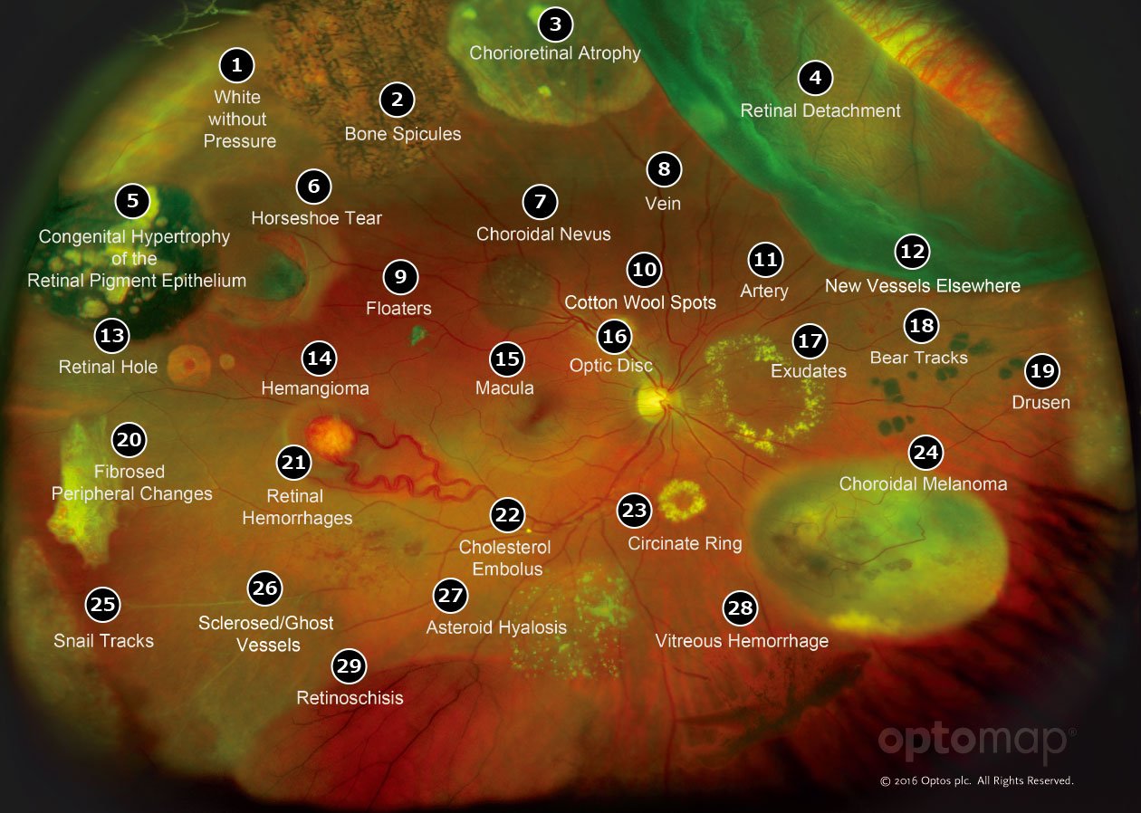
Diseases and other elements that can be detected through retinal imaging diagnostics
- 1:White without Pressure
- 2:Bone Spicules
- 3:Chorioretinal Atrophy
- 4:Retinal Detachment
- 5:Congenital Hypertrophy of the Retinal Pigment Epithelium
- 6:Horseshoe Tear
- 7:Choroidal Nevus
- 8:Vein
- 9:Floaters
- 10:Cotton Wool Spots
- 11:Artery
- 12:New Vessels Elsewhere
- 13:Retinal Hole
- 14:Hemangioma
- 15:Macula
- 16:Optic Disc
- 17:Exudates
- 18:Bear Tracks
- 19:Drusen
- 20:Fibrosed Peripheral Changes
- 21:Retinal Hemorrhages
- 22:Cholesterol Embolus
- 23:Circinate Ring
- 24:Choroidal Melanoma
- 25:Snail Tracks
- 26:Sclerosed/Ghost Vessels
- 27:Asteroid Hyalosis
- 28:Vitreous Hemorrhage
- 29:Retinoschisis