*This article introduces a project in Japan.

From making predictions about the global economy to smart device assistants, AI is greatly expanding its fields of activity. Nikon has been taking on the challenge of utilizing AI in the field of ophthalmology. A retinal imaging program that uses “deep learning” is one of the advances successfully achieved. This new AI-utilizing technology was jointly developed by DeepEyeVision* and Nikon.
- *DeepEyeVision is a startup company that emerged from Jichi Medical University in Tochigi, Japan in order to contribute its medical AI technology to society, established by assistant professor at the university Hidenori Takahashi (MD).
Jointly developed retinal imaging device program using AI
Along with the aging of society, eye diseases such as diabetic retinopathy and glaucoma, which can lead to blindness, also continue to increase. Against this background, early detection and appropriate treatment of these diseases are major factors in improving the quality of life for patients. Therefore, there was high demand for a medical device program that could provide ocular fundus imaging information to effectively support rapid diagnosis by ophthalmologists. Nikon and DeepEyeVision, utilizing deep learning technology, have jointly developed a program in which AI analyzes images captured by a retinal imaging camera. Their goal is to contribute to rapid and accurate diagnosis by ophthalmologists.
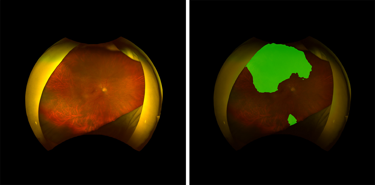
Right: Fundus image analyzed by AI
- *This is a program that further processes image information obtained from a fundus camera for use as an aid while diagnosing the health condition of a patient. It does not distinguish specific diseases nor diagnose the degree of progression of a medical condition, and does not include an automatic diagnosis function.
Improving the quality of medical care through helping both doctors and patients
An AI-powered analysis program for retinal imaging cameras has the potential to provide benefits to both ophthalmologists and patients. For doctors, it is expected to shorten the time required to “read*” an ocular fundus image. In addition, since it supports “reading” which has conventionally required physicians with long experience, this may make it easier for even less-experienced doctors to detect lesions.
For patients, the expected advantage is that lesions might be detected earlier. By using AI, we aim to improve the quality of medical care by helping both doctors and patients. Nikon continues to take on the challenge through its R&D of expanding AI technology that supports medical fields.
- *In this case, “reading” refers to obtaining medical findings from an examination image.