Quality of vision (such as eye health and clear view) is important for improving the quality of our lives. Nikon Eye Care Solutions assists the early diagnosis of eye disease including diabetic retinopathy that can lead to vision loss and visual field defect.
The core is the ultra-widefield retinal imaging device from the Nikon Group's Optos. This device captures an image of the fundus area with approximately 200˚ coverage simultaneously. The image contributes to early diagnosis of fundus lesions. The retinal images can be captured through 2mm pupils or non-mydriatic diagnosis, so the waiting time of approximately 30 minutes to 1 hour after applying instillation is eliminated. This approach is beneficial to patients because it allows them to return to everyday life right after inspection.
Optical system employing an ellipsoidal mirror
Optos ultra-widefield retinal imaging device uses a unique optical system with a concave three-dimensional ellipsoidal mirror to scan laser light across the retina. The ellipsoidal mirror reflects the laser light through the pupil to scan the retina. A galvanometer mirror capable of reciprocating oscillation at high speed, changes the angle of incidence of the laser beam during irradiation, enabling it to rapidly obtain high-definition images across an ultra-widefield of view.
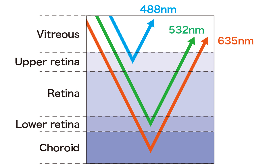
Using multiple laser lights for scanning
To view various depths of the retina, Optos technology incorporates two kinds of scanning laser (Red 635nm; Green 532nm) that cover different distances. The green laser scans from the sensory retina to the pigment epithelial layers, while the red laser scans from the RPE*1 to the choroid. Also, high-end equipment employs a blue scanning laser for fluorescein vascular inspections*2. In this way, lesion areas can be determined easily.
- *1RPE: Retinal pigment epithelium
- *2Fluorescein vascular inspections: Fluorescein is injected into vessels mainly for examining lesions to the retina and retinal pigment epithelium.