July 14, 2020
TOKYO — Nikon Corporation (Nikon) is pleased to announce the release of NIS-A Clarify.ai, an AI module for microscopes that uses artificial intelligence to remove signal emitted from defocused planes (out-of-focus image planes) from fluorescence images.
NIS-A Clarify.ai is a module for NIS-Elements Imaging Software that leverages deep learning, a type of artificial
intelligence. NIS-A Clarify.ai is pre-trained to recognize fluorescence signal emitted from out-of-focus planes and can remove this component from the image, resulting in significantly improved signal-to-noise (S/N) ratio.
Widefield, fluorescent microscopes offer multiple advantages over confocal systems, including fast acquisition speed, low phototoxicity, ease-of-use and low initial cost. A major drawback for widefield microscopes has been the contribution of out-of-focus light and resulting degradation of S/N ratio. NIS-A Clarify.ai enables users to easily acquire high-contrast, high S/N ratio images with widefield fluorescent microscopes. In addition, quantitative analysis is possible by combining a wide variety of image analysis functions in NIS-Elements software.
NIS-A Clarify.ai enables clear visualization of biological phenomena in live samples, even those occurring in deep areas, providing an important contribution to various fields of research including cell biology, neurobiology and embryology. It also improves workflow efficiency through simple processing.
Product Overview
| Name of Product | “NIS-A Clarify.ai” AI module for microscopes |
|---|
Major Features
1. Improves sharpness of fluorescence images with one click
When focusing on an object to acquire fluorescence images, image clarity may be impaired by out-of-focus signal emitted from above and below the focal plane. Traditionally, users have relied on deconvolution to remove out-of-focus light from widefield fluorescence images. NIS-A Clarify.ai is pre-trained to recognize out-of-focus signal and can remove this component from widefield fluorescence images. An advantage of NIS-A Clarify.ai compared to deconvolution is fast image processing.
The pre-trained NIS-A Clarify.ai removes fluorescence signal emitted from out-of-focus planes with one click thus rapidly increasing image contrast and reducing research time and workload.

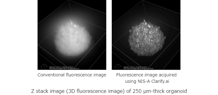
2. Create high-definition 3D images from widefield fluorescence images
By stacking multiple 2D fluorescence images processed with NIS-A Clarify.ai, high-definition 3D fluorescence images can
be created.
This new AI-based module allows high-contrast images to be acquired even in deep areas of a thick, live sample, and is
expected to be applied to morphological research. Click to watch the following sample movie.
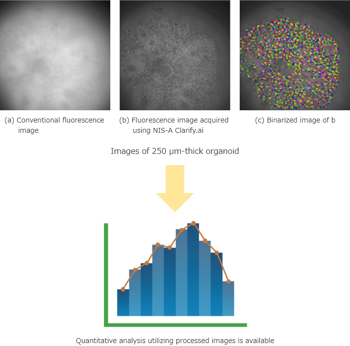
3. Allows quantitative analysis through seamless linkage with image analysis functions
Images processed using NIS-A Clarify.ai can be quantitatively analyzed using a wide variety of image analysis functions of the NIS-Elements software, allowing easy analysis of research results.
The information is current as of the date of publication. It is subject to change without notice.