March 18, 2025
Nikon Corporation (Nikon) has developed ECLIPSE Ui version 1.4, a software update to version 1.4 for its digital imaging microscope. The update will be phased in starting from March 18 into ECLIPSE Ui models sold by Nikon and its local distributors.
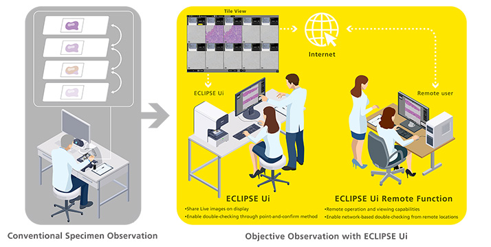
The ECLIPSE Ui medical device continues to improve pathological observation workflows with its unique, eyepiece-less design and remote control and viewing functions via network.
The update includes new functions to streamline the pathology observation workflow, including Tile View and Layer View, which can record up to 20 specimen images consecutively and allow up to 10 specimen images to be compared on a single screen. In addition, the Remote Mode function, which supports data viewing in remote locations, has been enhanced. This makes data sharing with doctors and teams in remote locations more efficient and smoother, further reducing the burden on pathologists and clinical sites.
Development Background
The domestic clinical testing market is facing a shortage of pathologists. As of October 2024, there were only 2,841 doctors specializing in pathology observation in Japan[1], resulting in a high observation burden per person. For this reason, there is a demand for techniques that can achieve efficient and accurate observation with limited resources.
Pathology tests to observe diseases such as cancer use a variety of staining methods, including "HE staining" to observe the shape of tissue, "immunostaining" which uses antigen-antibody reactions, and "special staining" which uses chemical reactions.
Previously, it was necessary to slice multiple specimens from the same tissue and stain them with different dyes before observing them. In many cases, multiple dyes were required to make a single observation, and the task of finding the same area in each specimen had to be repeated, placing a burden on pathologists.
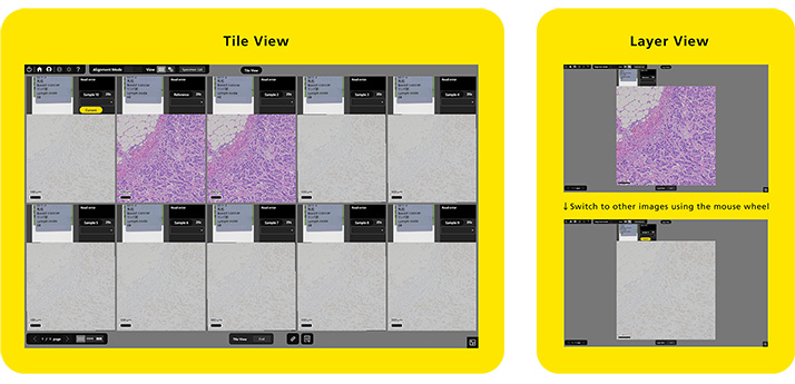
To address these issues, Nikon developed the Alignment Mode for its ECLIPSE Ui Digital Imaging Microscope. Version 1.4 includes two new functions: Tile View, which records up to 20 specimen images in succession and displays up to 10 images on one screen, and Layer View, which switches between images of the same location stained with different methods. The new functions make it easier to compare images and eliminate the need to switch specimens.
Additionally, clinical sites face challenges such as regional disparities, aging medical practitioners due to a shortage of pathologists, fatigue from long periods of observation and coping with difficult cases. Given these conditions, the update includes functions to enhance remote observation efficiency and support seamless teamwork.
Nikon has developed ECLIPSE Ui Version 1.4 as a observation support tool that integrates microscope and digital technologies. This enables fast and accurate observation with limited medical resources, helping to address challenges in pathology observation and improve overall efficiency in medical treatment environments.
- 1.Number of pathologists certified by the Japanese Society of Pathology
Release Overview
| Product Name | ECLIPSE Ui Digital Imaging Microscope |
|---|---|
| Version | 1.4 |
| Availability Start Date | Initial roll out beginning March 18, 2025 |
| Key Updates |
|
Main Update Features
1. High-quality overall display and digital zoom functionality enable quick access to lesion sites, supporting improved observation efficiency.
In Tile View, multiple images captured at the same location are displayed in parallel, making it easy to compare differences between stained images. In Layer View, multiple images of the same location can be switched seamlessly, enabling comparison of HE-stained tissue morphology with antigen localization in immunostaining. This reduces the effort required to locate the same region across different specimens, a challenge in conventional observation workflows, thereby supporting improved observation efficiency.
2. Enhanced Remote Operation for More Efficient Telemedicine Support
Features introduced in previous versions are now available for remote observation, enabling smoother and more efficient data sharing with physicians and teams in different locations.
Key Features:
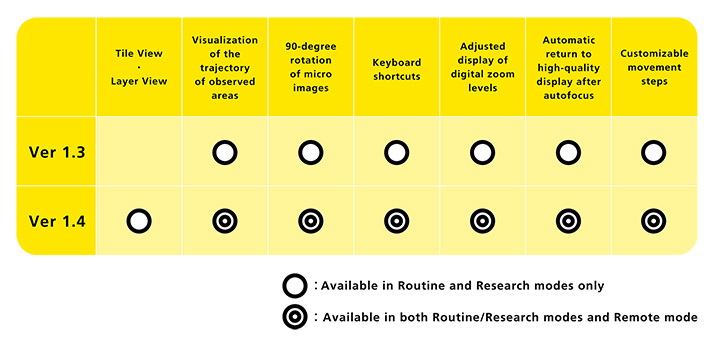
- Visualization of the trajectory of observed areas: Displays examined fields on macro images to help prevent oversight errors.
- Customizable movement steps: Enables systematic examination based on observation conditions.
- Enhanced usability:
- Expanded keyboard shortcuts
- Automatic return to high-quality display after autofocus
- Adjusted display of digital zoom levels
- 90-degree rotation of micro images
These enhancements improve remote observation efficiency and facilitate seamless collaboration across locations.
The information is current as of the date of publication. It is subject to change without notice.