
November 12, 2026
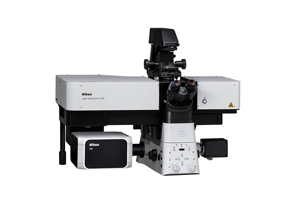
Nikon Corporation today announced the LS-X Light Sheet Unit, designed to capture clear images within three-dimensional specimens, from living organoids and embryos to cleared samples and specimens prepared using expansion microscopy. Built around Nikon’s ECLIPSE Ti2-E inverted research microscope, the LS-X supports a wide range of magnifications and research workflows, including gentle live imaging. Phased availability is scheduled to begin from November 30, 2026, with a gradual launch across worldwide regions through Nikon and its local distributors.
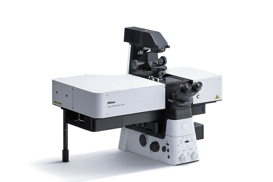
Supporting a broad range of 3D research
Organoids are three-dimensional cell cultures that reproduce selected features of human tissues. They are increasingly important in drug discovery and regenerative medicine, where researchers use them to investigate biological processes and disease. Organoids derived from patient cells also offer opportunities to study differences between individuals and support the development of personalized approaches to treatment.
These models change over time. Following their development and internal activity requires imaging methods that can reveal detail within a specimen while preserving its viability throughout an experiment.
Other approaches help researchers examine the organization of biological structures in prepared samples.
Tissue clearing reduces light scattering, while expansion microscopy physically enlarges specimens to make fine structures easier to resolve. Together, these applications create a need for flexible imaging across different specimen preparations and scales.
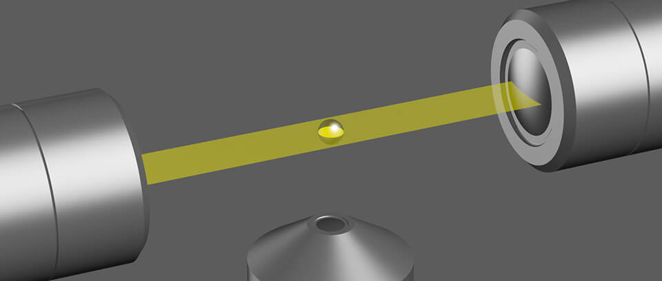
Light sheet microscopy addresses this need by illuminating a thin plane within the specimen and capturing the resulting fluorescence with a camera. Restricting excitation to the region being imaged helps reduce background and unnecessary light exposure. The LS-X brings this approach to Nikon’s inverted microscope platform, with automated alignment and dedicated sample chambers designed to simplify preparation.
Product launch overview
| Product name | LS-X Light Sheet Unit |
|---|---|
| Release date | Phased Availability Beginning November 30, 2026* |
- *Availability dates may vary by region.
Key features
1. Clear imaging within three-dimensional specimens
The LS-X illuminates specimens from two sides to support imaging within living samples, cleared specimens, and samples prepared using expansion microscopy. By concentrating excitation around the plane being observed, light sheet illumination reduces out-of-focus background and helps reveal structures within the specimen.
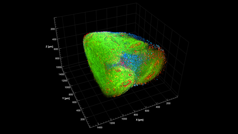
2. Gentle Live Imaging for Delicate Specimens
Selective illumination helps reduce light exposure during repeated imaging, supporting time-lapse studies of living specimens. The LS-X also supports near-infrared excitation.
A dedicated LS-X incubator manufactured by Tokai Hit Co., Ltd. supports extended live-imaging experiments.
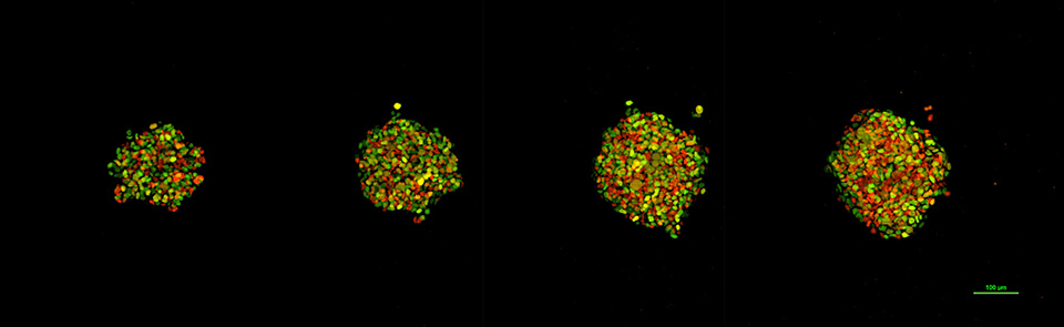
Cells are labeled with the Fucci fluorescent reporter, which visualizes cell-cycle progression by color-coding cells in green, red, and yellow.
3. A wide range of magnifications and imaging techniques
The LS-X supports a wide range of magnifications, allowing researchers to move from broader views of a specimen to closer examination of selected structures. This flexibility supports imaging across different sample sizes and research needs.
The flexibility of the ECLIPSE Ti2-E base platform also enables workflows using various microscopy techniques, including light sheet, confocal, and epifluorescence imaging. Researchers can select complementary methods within a shared microscope platform. For compatible existing Ti2-E systems, the LS-X offers a way to add light sheet imaging to an established microscopy platform.
4. Simpler preparation and alignment
Nikon’s NIS-Elements imaging software is designed to automate the optical alignment required for light sheet microscopy, reducing manual setup.
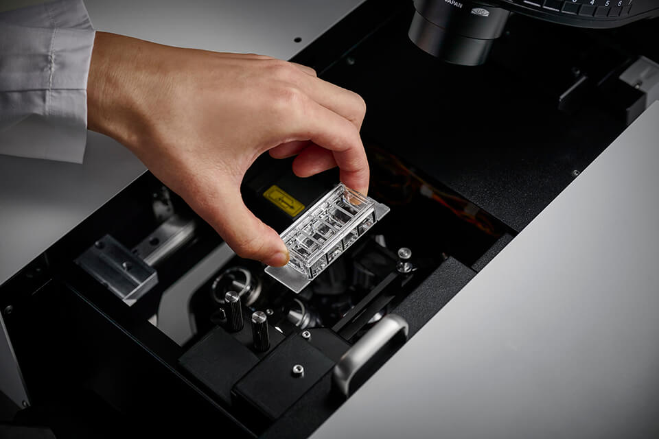
Dedicated LS-X multi-chamber slides support workflows from cell culture and embedding through imaging without transferring the specimen to another vessel. The chambers are designed for light sheet imaging and also support confocal and epifluorescence microscopy, helping researchers use complementary methods with the same sample.
Comment from Haruhisa Sonoda, Head of Nikon Healthcare Business Unit
“Researchers study biology at many scales, from changes in living three-dimensional models to fine structures within prepared specimens,” said Haruhisa Sonoda, Head of Nikon’s Healthcare Business Unit. “The LS-X brings light sheet imaging to the flexible Ti2-E platform, giving researchers more ways to approach these questions. We want to help them spend less time preparing the microscope and more time investigating the biology. Nikon will continue to draw on its optical and microscopy expertise to support advances in life science research, drug discovery, and regenerative medicine.”
About Nikon’s Healthcare Business
Established in 2017, Nikon’s Healthcare Business brings together expertise in biological microscopy, medical imaging, and cell manufacturing. It operates in three areas: Life Science Solutions, Eye Care Solutions, and Contract Cell Manufacturing Solutions.
Life Science Solutions provides microscopes and imaging technologies that help researchers study the structures and behavior of living cells. Its offerings include super-resolution microscopy, cell culture observation systems, and cell-related services for research in regenerative medicine and drug discovery.
Eye Care Solutions provides ultra-widefield retinal imaging technologies that support the detection and treatment of eye disease.
Contract Cell Manufacturing Solutions provides pharmaceutical companies and biotechnology ventures with cell development and manufacturing services for regenerative medicine and gene therapy.
Through these activities, Nikon’s Healthcare Business works to advance research and healthcare and improve quality of life.
Related Link
Nikon Healthcare biological microscope products and services website
The information is current as of the date of publication. It is subject to change without notice.