
Have you ever heard of “time-lapse photography”? It’s a shooting method that captures a series of frames at time intervals of seconds, minutes, tens of minutes, or even a day, and compresses the changes occurring over a long period of time into a short-length movie. Yone Production Co., Ltd. has been implementing time-lapse photography using microscopes for more than half a century – with continued success in capturing images of living cells and bacteria. Recently, at the request of a TV broadcaster, they succeeded in filming how the COVID-19 virus destroys cells. Find out how Nikon microscopes are used for these unique filming techniques.
Time-lapse photography that visualizes movements of cells and bacteria.
In 1958, a scientific movie produced in Japan won the Best Scientific Film Award at the Venice Documentary Film Festival along with other accolades at domestic and international film festivals. The film was titled ‘The World of Microbes – In Quest of the Tubercle Bacilli’ and it employed time-lapse photography* to depict the battle between white blood cells and tubercle bacillus, the cause of pulmonary tuberculosis, for which no effective treatment had yet been found. This film astonished the world with its groundbreaking technical results that clearly captured the movement of cells (white blood cells) and bacteria that had never previously been seen in a movie.
We interviewed Ms. Ayumi Fujieda, CEO of Yone Production Inc., who explains. “Yone Production Co., Ltd. was established in 1967 by Mr. Yonesaku Kobayashi, a photographer who played a central role in the production of the ‘The World of Microbes’ film, along with Mr. Tokio Asaka who is still currently active within the company to this day.”
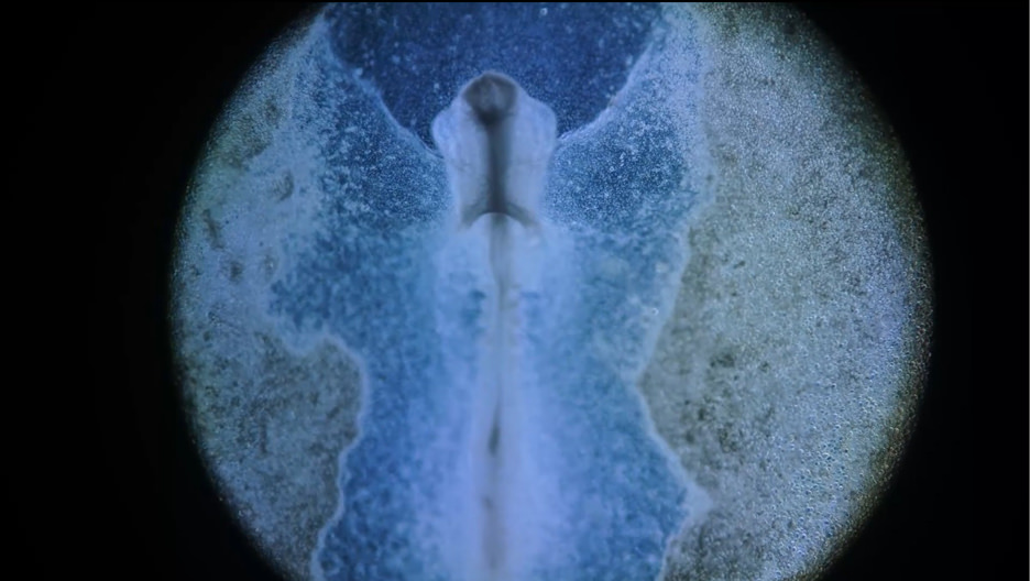
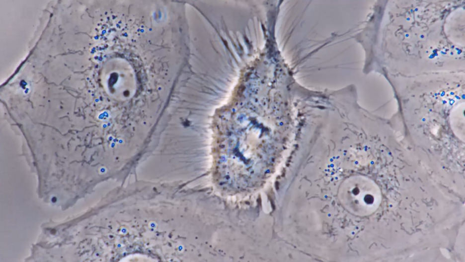
Since its inception, Yone Production Co., Ltd. has continued through the years to photograph cells, biological tissue, bacteria, and microorganisms employing its own unique technology – becoming one of the world’s leading companies in the field of time-lapse photography using biological microscopes. Technology capable of visualizing the invisible world has played a huge role in contributing to the fields of medicine, medical care, and drug research. Ms. Fujieda also explained that a recent project involved 8K filming of cells infected with COVID-19, which was conducted at the request of a TV station under the guidance of the Research Institute for Microbial Diseases, Osaka University.
This video was the first in the world to capture the destruction of cells by a virus, which not only attracted the attention of many scholars and medical experts, but also garnered considerable interest from the general public as well.
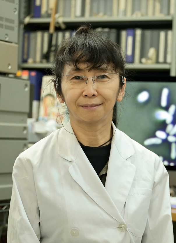
CEO of Yone Production Co., Ltd.
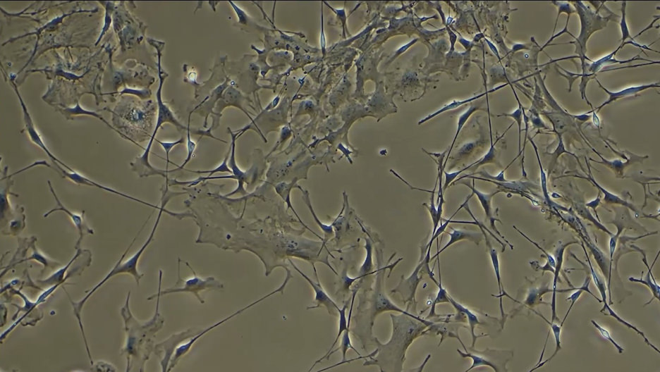
From the movie “The universe in the body” in 2018.
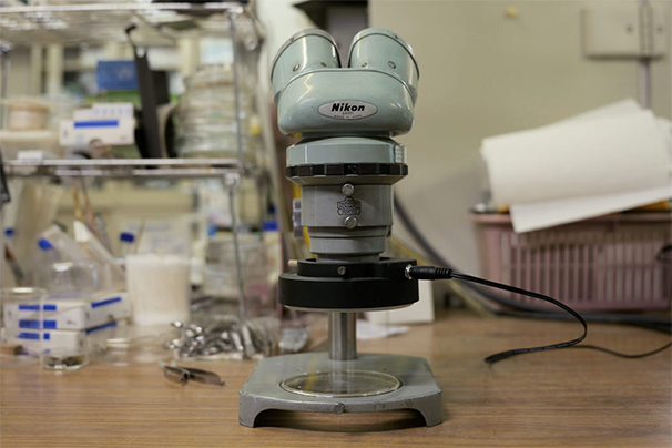
*Nikon MICROPHOT was used.
From the movie “The universe in the body” in 2018.
*Nikon DIAPHOT TMD was used.

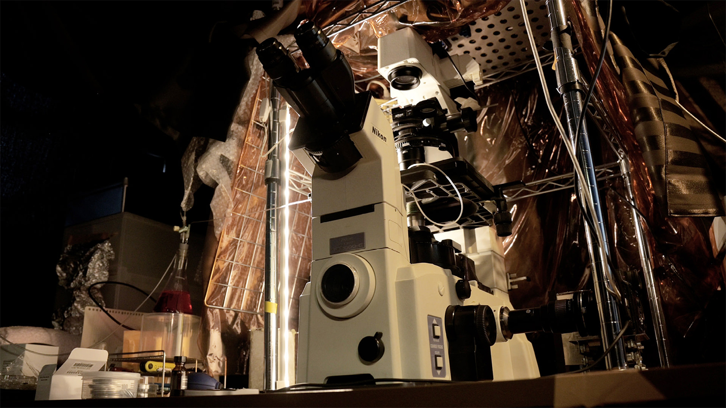
From the movie “The universe in the body” in 2018. *Nikon ECLIPSE Ti2 was used.
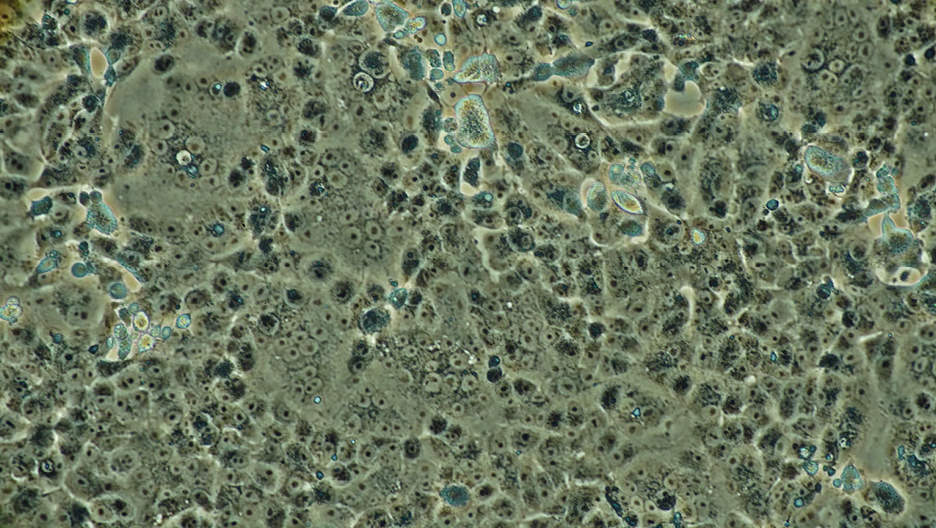
Photo courtesy: Osaka University
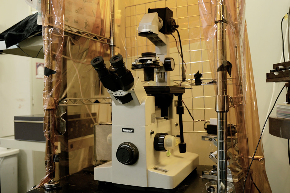
*Nikon ECLIPSE TE2000 was used.
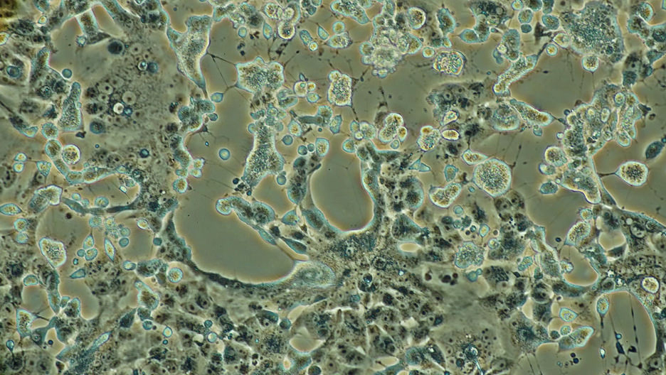
Photo courtesy: Osaka University
*Nikon ECLIPSE TE2000 was used.
- *Time-lapse photography captures images at preset intervals. By connecting still images after shooting, slow changes over a long period of time can be reproduced as a short film.
Films created by the synergy of researchers and photographers
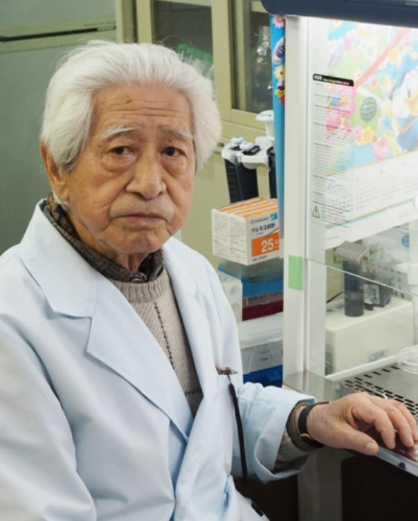
Yone Production Co., Ltd. has a production department, directing department, photography department, and a research department that produces biological samples. The research department first reads through and examines materials and papers from clients such as academic institutions and societies, government ministries and pharmaceutical companies and shares the relevant content with the production and directing departments. The research department is responsible for thinking about exactly how to cultivate the subjects to be observed and how to prepare them in a living state in petri dishes according to the plan that takes shape following a briefing. Mr. Tokio Asaka, introduced by Ms. Fujieda earlier, said, “We need to present specimens in such a way as to capture clear, innovative, and academically convincing images. This is extremely difficult.” He added, “I sometimes have disagreements with the photography department, but this can also tend to help us make discoveries which advance the project. I think this is the Yone Production way.” Born in 1925, Mr. Asaka is now 96 years old and still active in the company.
Director, Head of the Academic Division of Yone
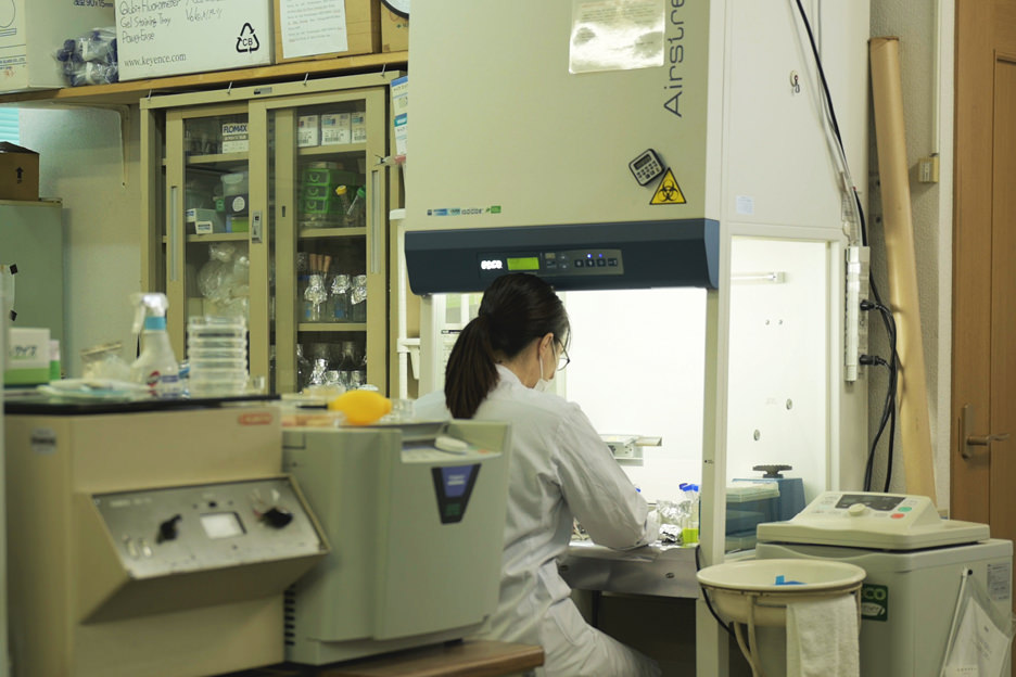
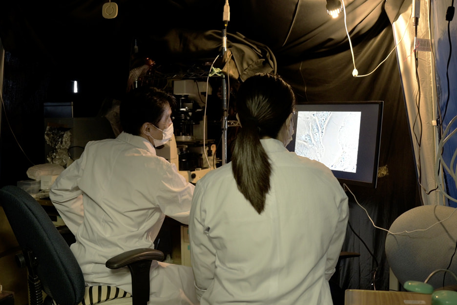
Next, we interviewed Mr. Hiromi Fujimoto from the photography department. He is a specialist who has worked on numerous projects and is one of a rare breed of photographers working with a scientific perspective. He was in charge of shooting the 8K movie of COVID-19 previously mentioned and tells us, “The most important thing in time-lapse photography using microscopes is having a clear aim of what kind of subject and what kind of phenomenon to shoot – but most of the time this doesn’t go exactly as planned. We’ve been going through a careful trial and error process on how to solve what doesn't work. I think that determination plays a big part in our success.”
Currently, Yone Production Co., Ltd. consists of a small team of six people. It is a group of dedicated life science film specialists who have research functions that ordinary film production companies do not, and all the staff members share their collective knowledge and experiences. These are some of the reasons why they can continue to produce images that amaze the world.
MicroCinematographer
of Yone Production Co., Ltd.
Nikon’s biological microscopes respond to the experimentation and ingenuity of specialists.
Yone Production Co., Ltd. consistently strives to create new images using the special techniques of micro-mirror time-lapse photography. The company’s equipment allows for ample trial and error along with the team’s groundbreaking ingenuity. Unique, tried and tested ways of using microscopes have been taught by previous researchers since the company was founded.
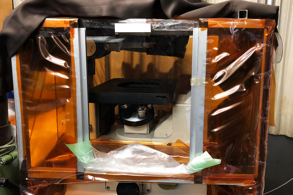
For example, in order to keep cells viable for extended periods of time, the areas around microscope stages are processed to maintain certain temperatures and humidity levels – achieved by pumping in CO2. The team has employed special petri dishes, made boxes from materials purchased at DIY stores, and procured parts from electronics stores. Subsequently, various manufacturers put their ideas into practical use and these were later commercialized as devices such as chambers for microscopes*1. It can be said that such advanced equipment was created based on their unique needs.
A recent example is the use of condensers as a light source. For shooting subjects with a certain thickness, an upright microscope*2 is customized to use the condenser of an inverted microscope*3, or the light source is moved as far as possible to one side.
Mr. Hiromi Fujimoto, Technical Director/MicroCinematographer of the filming department, remarks, “I have used various microscopes, but Nikon microscopes work without the stage rattling or going out of focus. You can take any kind of image you want.” Many Nikon microscopes, including the ECLIPSE TE2000, are located in the research and shooting departments. Some of the microscopes are more than 50 years old and still in use.
These are true specialists who work to uncover the secrets of cells and bacteria using unique methods, visualizing them and contributing to the ongoing development of medical and life sciences. It is a great honor for Nikon’s microscopes to proudly play a part in their achievements.
- *1Microscope chambers are devices to keep the temperature and humidity levels on the stage constant and enable the cultivation and observation of cells.
- *2An upright microscope is a microscope that has an objective lens above the sample to be observed. The condenser (light source) is located on the lower side of the stage.
- *3An inverted microscope is a microscope that has an objective lens located under the sample to be observed. The condenser (light source) is located on the upper side of the stage.