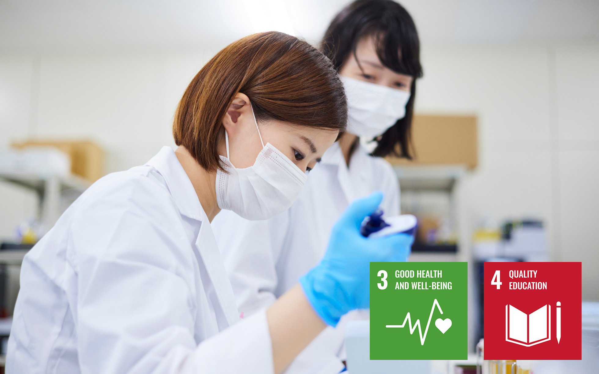
Healthcare plays an extremely important role in the enhancement of QOL (quality of life). In Japan, with a declining birthrate, aging population, growing awareness of healthcare and advances in medical technology, medical technologists that possess specialized knowledge and skills for clinical laboratories are in great demand in order to support physicians. Saitama Medical University Faculty of Health and Medical Care is raising the next generation of such healthcare professionals. At the School of Medical Technology, Nikon’s microscopes are being used to help students during their clinical laboratory training.
Training medical technologists to support physicians.
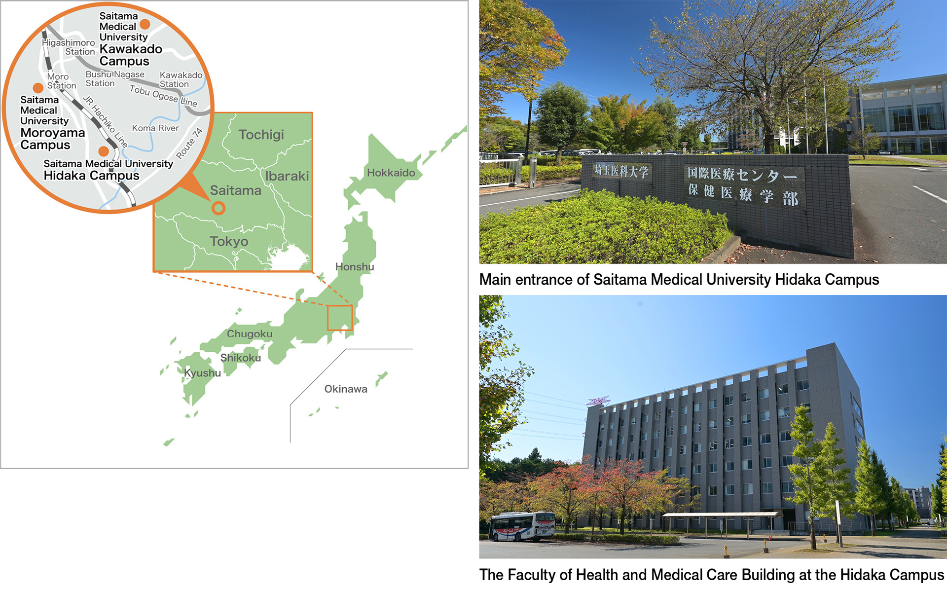
Saitama Medical University Faculty of Health and Medical Care was established in 2006, with the aim of raising a new generation of professionals with healthcare and medical knowledge and skills, in order to contribute to peoples’ health and welfare in this new era of medical care.
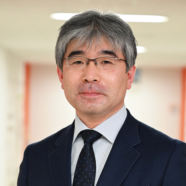
Here, we talk to Professor Tsuyoshi Onogawa, who explains “The Faculty of Health and Medical Care currently includes four departments, the School of Nursing, the School of Medical Technology, the School of Bioengineering, and the School of Physical Therapy. I work as a professor at the School of Medical Technology.” He goes on to say that “The School of Medical Technology is a department that trains medical technologists. Students learn about the mechanisms of diseases by studying, for example, physiology, pathology and immunology, and based on these contents, they also get to know how to assist in diagnosis through lectures and practical training. The students will also need to study public health and nutrition, plus cutting-edge areas such as genetic testing. As the scope of this learning process is so extensive, it is said that it is comparable to that of medical schools for doctors, minus the actual treatment part.”
Faculty of Health and Medical Care, School of Medical Technology, Immunology
Professor Tsuyoshi Onogawa
Together with medical technology, clinical laboratory technology is also evolving every day. By means of improvements in the accuracy of various testing equipment, development of new testing methods, and the high skills of medical technologists, more accurate and rapid testing is made possible, thereby supporting physicians’ diagnosis and treatment. Moreover, this contributes to the maintenance and management of health through health checkups and other examinations. These are techniques and skills that are essential to improving the QOL of many people.
The microscope: Partner of medical technologists.
The microscope is an indispensable instrument in the process of learning a wide range of medical knowledge and acquiring the techniques and skills of a specialist, through lectures and practical training in various examinations. Professor Onogawa comments that “The microscope is the first piece of examination equipment that students aiming to become medical technologist will be using. They will build an enduring relationship throughout their technologist career with microscopes, as not only will they be using them while in school, but also when engaged in examinations. In other words, microscopes are their essential partners.”
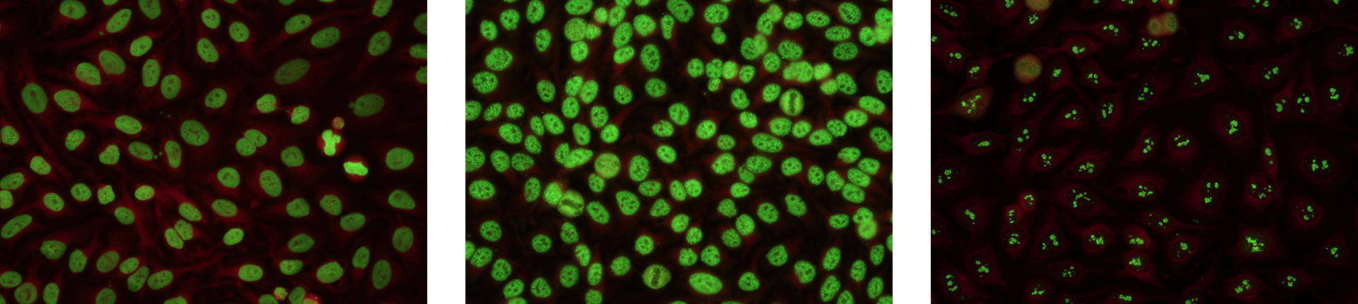
Practical training with microscopes ranges from histology and pathology to blood and microbiology specimens. Among them, we had the opportunity to learn about the clinical immunology training that uses Nikon’s fluorescence microscope*1, the ECLIPSE Ci-L. The ‘antinuclear antibody test’ studied here is frequently utilized for exam photos in the national examination for medical technologists. However, a single fluorescence microscope is often shared by many students in a lot of educational institutions, and in such cases, each person can only observe for a very short time. “I’ve always wished that each student could spend more time making closer and more detailed observations that they will be able to remember more clearly. It doesn’t really matter how beautiful an image shown on a screen might be, as it looks and feels completely different from the actual fluorescent image observed through an eyepiece”, the professor comments. This wish came true at Saitama Medical University. “We discussed with Nikon about the possibility of arranging fluorescent microscopes for each training group that consists of around five members, and they agreed to lend us the necessary number of microscopes. This allowed us to increase the amount of time each student could spend making observations. We also had the opportunity to arrange for Nikon people to visit and explain about the mechanism of the fluorescent microscopes to the students, so that they could gain a fuller understanding regarding fluorescent images. Such images are very impressive when you see them in real life, and most students that see them for the first time through the fluorescent microscopes are in awe at how amazing how they are. And I was actually one of those students back in the day,” says Professor Onogawa, “so we are very grateful to be able to maintain such a training environment.”
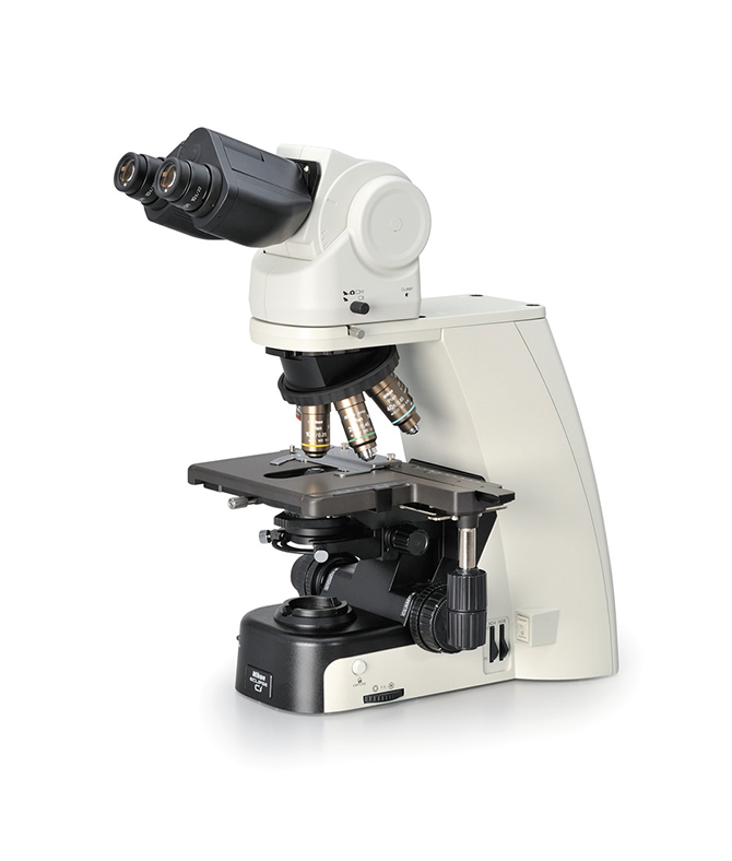
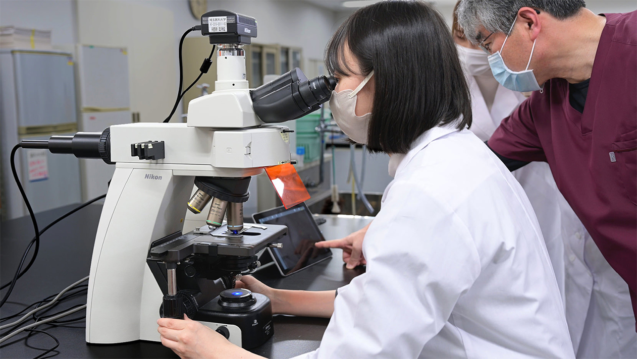
When we asked him what the main reasons were behind deciding to use the ECLIPSE Ci-L, he explained that the microscope itself plus the required additional light source unit, are overall very compact, the combination provides high resolution and is extremely bright even when performing bright field observation*2, moreover, the attractive design was another of the decisive factors.
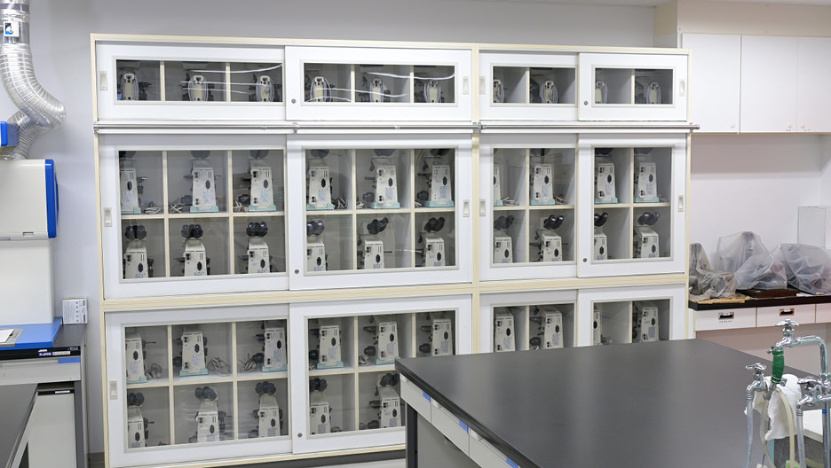
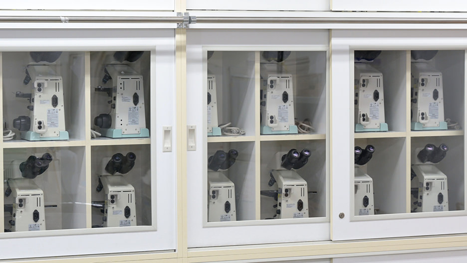
Other than the ECLIPSE Ci-L, the students are also employing Nikon’s ECLIPSE E100 microscopes for their practical training at the university. There are more than 80 sets of these microscopes, used by not only the School of Medical Technology, but also the School of Nursing and the School of Physical Therapy for their practical training. There are also custom-built cabinets with glass doors for storing the microscopes, allowing anyone to quickly check that they are properly managed. All this shows how medical technologists take great care of and value their equipment.
- *1Fluorescence microscopes are instruments employing a special light source that can excite (illuminate) fluorescent biological specimens.
- *2Bright field observation is an observation method in which samples such as pathology specimens and blood samples are observed using a transillumination or other light source.
The human resources of tomorrow and the future of healthcare.
Lastly, we asked Professor Onogawa about his thoughts on human resources development. He answered that “It’s important to learn the basics well and never neglect the timeless principles. I also hope that students will always remember to be considerate of others.” He hopes that not only will they acquire the knowledge, techniques, and essential skills, but also prepare themselves mentally to become a valued part of the medical community.
Professor Onogawa is currently involved in research regarding sepsis, an important subject in the field of emergency medicine. He emphatically states that, “My dream is to be able to unravel how we can avoid acute organ failure caused after infections through immunological approaches, and provide emergency doctors who are making their best efforts in saving lives with a mindset that serves as the basis of new treatment methods. We are constantly striving with students to help them not only develop into medical technologists, but also as individuals with a mindset for making research by having this as their senior thesis topic. The microscope is my indispensable partner for this research.”
Committed to fostering the medical professionals of tomorrow and pursuing the possibilities of future healthcare, Nikon microscopes will continue to contribute to these goals.