- *This page contains interviews with healthcare professionals regarding our products. However, these interviews do not guarantee the efficacy, effectiveness, or performance of the products, nor do they constitute an official endorsement, recommendation, advice, or selection by the featured professionals.
The declining birth rate, driven by lifestyle changes, is a critical social challenge that demands attention. Advancements in assisted reproductive technology (ART) are further anticipated to play a key role in addressing this issue. Fujita Medical Innovation Center Tokyo (FMiC) is committed to clinical practice, research, and development in ART to help more people wishing to have children and become parents. Nikon microscopes play a valuable role in supporting these efforts.
A new hub for advancing assisted reproductive technology
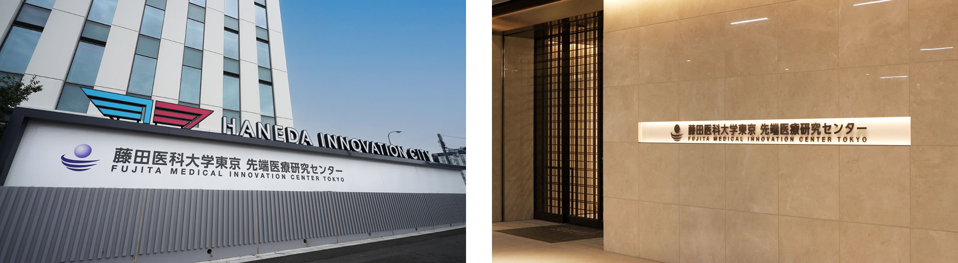
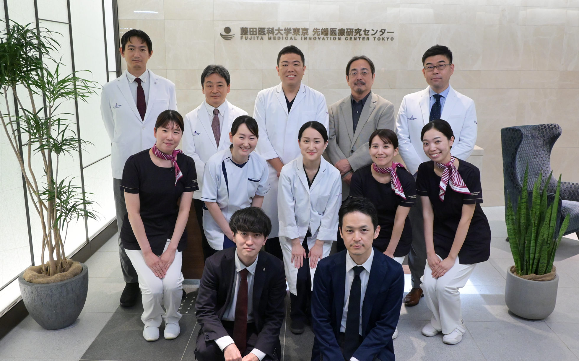
Fujita Medical Innovation Center Tokyo
The Center for Advanced Reproductive Medicine
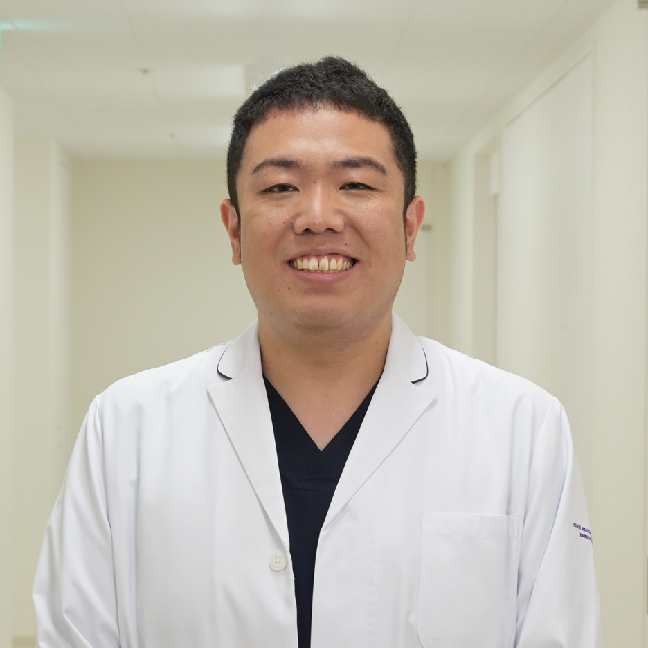
Department of Regulatory Science
Associate Professor
Tatsuya Kobayashi, Ph.D.
*Position and affiliation mentioned are as of the time of the interview.
Assisted reproductive technology (ART) encompasses infertility treatments such as in vitro fertilization (IVF). In Japan, approximately 8.6%*1 of all infants are now born through the use of ART, while in the United States, about 2.6%*2 are conceived in this way. We recently visited the FMiC to learn more.
Established in 2023 by Fujita Health University — a comprehensive medical institution with over 50 years of history — the center was created to address the evolving needs of next-generation healthcare. Located conveniently at Haneda Innovation City, directly connected to Tenkubashi Station and near Haneda International Airport, the center is actively advancing research in areas such as regenerative medicine and cancer genomics. In particular, it is deeply involved in advancing ART through clinical practice, research, and the training of future medical professionals.
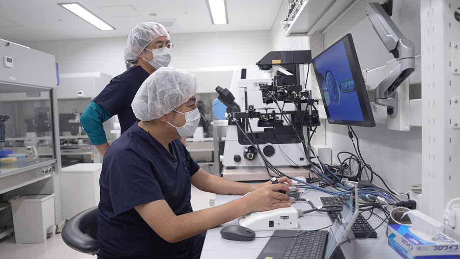
“I work as an embroyologist at the Fujita Health University Haneda Clinic, which is part of the center. Embryologists are responsible for procedures such as in vitro fertilization (IVF) and embryo cultivation under the supervision of physicians,” explains Associate Professor Dr. Tatsuya Kobayashi from the Department of Regulatory Science at Fujita Health Univeristy. “At the clinic’s embryo incubation room, we carefully optimize each step of the process — from selecting culture media, which significantly impacts the success of in vitro cultivation, to fertilization, embryo culture, freezing, and thawing for transfer. Our aim is to help patients achieve pregnancy as efficiently as possible,” he shares.
The FMiC and Haneda Clinic utilize a range of advanced tools and technologies to support the work of embryologists and physicians in both research and clinical applications of ART. Among these, Nikon microscopes play a vital role in their efforts.
- *1According to data published by the Government Public Relations Office of Japan in 2023
(https://www.gov-online.go.jp/useful/article/202309/2.html![[別窓で遷移します]](/common/images/icon/icon_blank.svg) )
) - *2According to 2022 data published by the U.S. Centers for Disease Control and Prevention
(https://www.cdc.gov/art/php/national-summary/index.html?cove-tab=2, https://www.cdc.gov/nchs/fastats/births.htm)
Microscopes advancing clinical practice and research
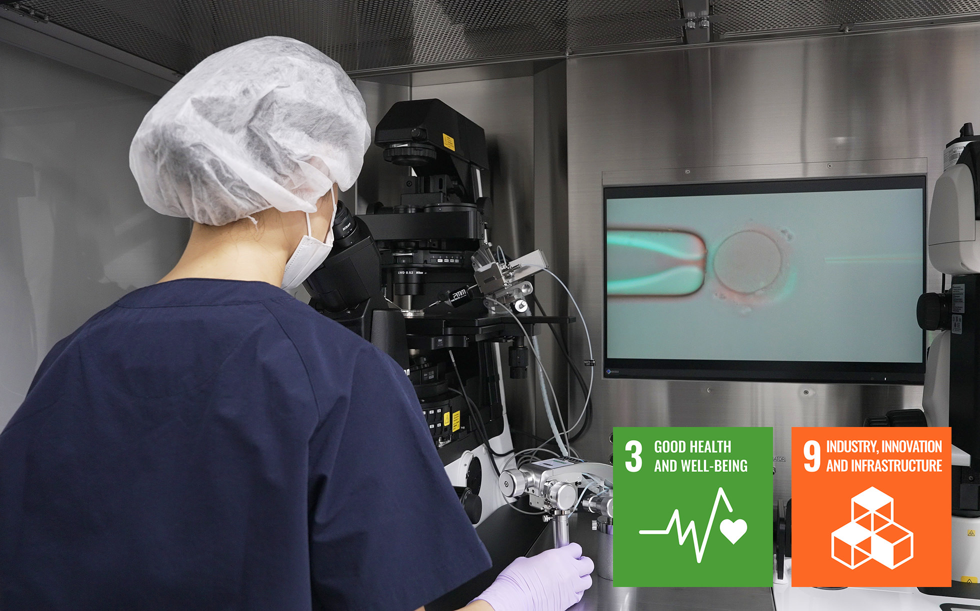
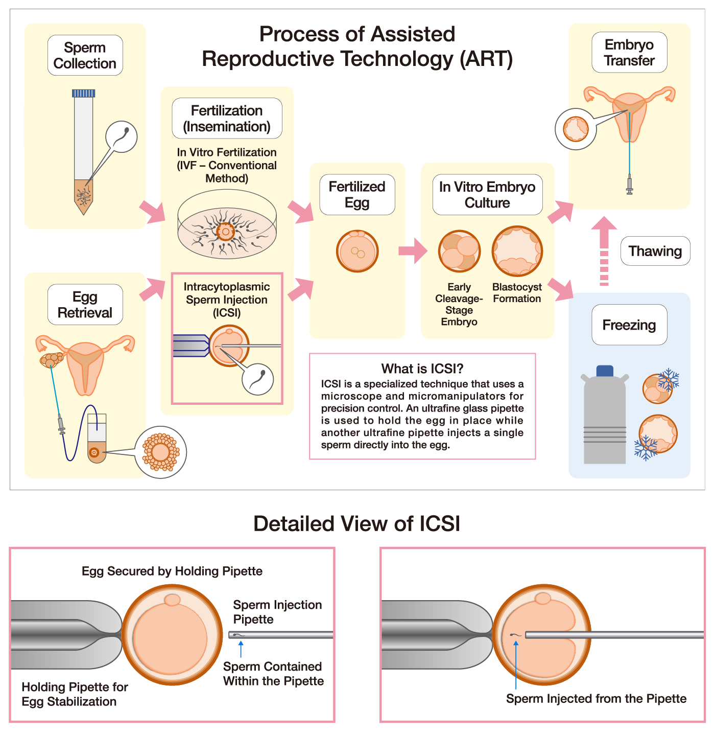
ART involves fertilizing eggs with sperm outside the body, culturing the resulting embryos, and transferring them to the uterus. There are two primary methods of fertilization. Conventional IVF involves adding sperms to a culture medium containing the egg, allowing natural fertilization to occur. On the other hand, intracytoplasmic sperm injection (ICSI) directly injects a single sperm into an egg using an ultrafine glass pipette. Once fertilized, embryos are cultured in an environment that mimics the human body, progressing through cell division to the blastocyst stage, and are then transferred to the uterus based on the patient’s condition.
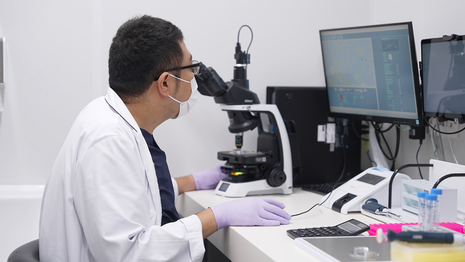
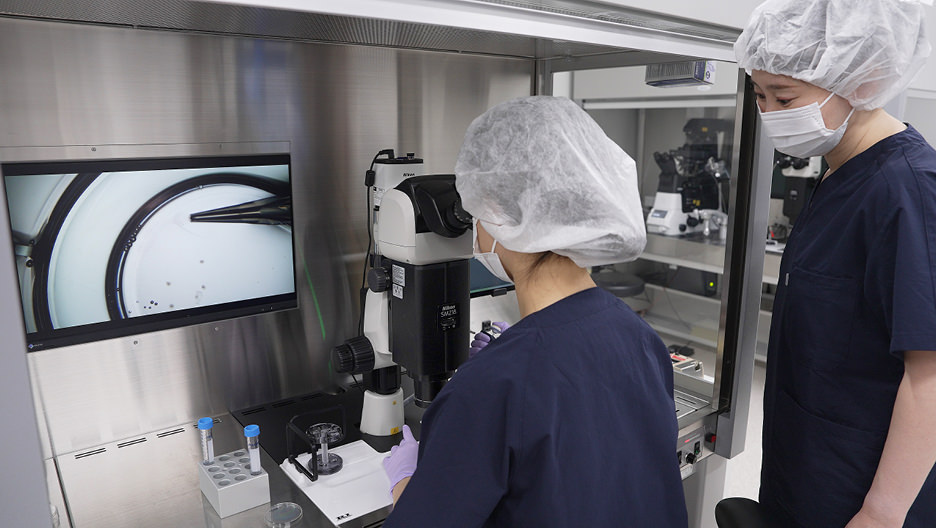
Microscopes are essential tools during the many steps of ART. At FMiC, Nikon’s stereo microscope, the SMZ18, is used to locate eggs within follicular fluid retrieved from the ovaries. Sperm samples are analyzed for motility and condition using Nikon’s upright microscope, the ECLIPSE Ci. The center also utilizes an automated sperm analysis system that incorporates the ECLIPSE Si upright microscope.
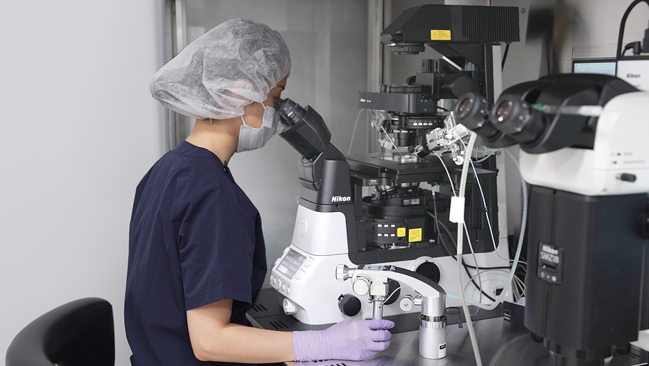
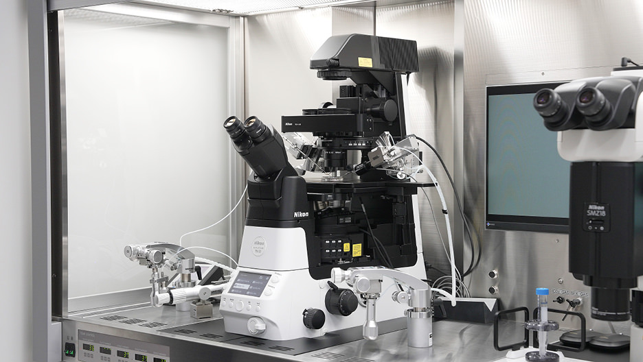
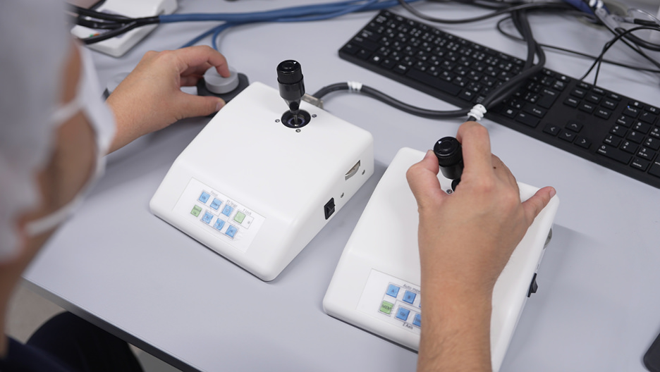
Microscopes play a particularly important role during fertilization. For ICSI, the ECLIPSE Ti2-I is involved in securing the egg, aspirating the sperm, and injecting it into the egg using ultrafine glass pipettes or needles precisely sized for the procedure. The ECLIPSE Ti2-I offers advanced features, such as objective lenses capable of displaying the egg’s nucleus (spindle) in color and high-magnification lenses for detailed sperm observation. These lenses require specific filters, which the Ti2-I selects automatically, streamlining the process and reducing the workload for embryologists.
Microscopes are also pivotal in research efforts. “We use Nikon’s ECLIPSE Ti2-I to conduct studies for confirming the improvement of workflow efficiency for embryologists involved in ART,” says Dr. Kobayashi. “The number of ART procedures is growing worldwide, but the shortage of embryologists remains a significant challenge. In our studies, we compared the time required for ICSI using the automated ECLIPSE Ti2-I versus conventional fully manual microscopes and the results proved that there was a noticeable reduction in procedure time. Moving forward, we plan to quantify workload metrics and continue implementing methods to support embryologists,” Dr. Kobayashi explains.
Advancing the potential of ART

Reproductive Engineering Laboratory, Laboratory Head
Kanto Branch, Director
Tomoo Eto, Ph.D.
*Position and affiliation mentioned are as of the time of the interview.
When discussing future challenges, Dr. Kobayashi explained: “We are collaborating with Dr. Eto from the Central Institute for Experimental Medicine and Life Science (CIEM) to develop microscopes and micromanipulators* based on Nikon’s ECLIPSE Ti2-U, that are capable of remote operation. This initiative aims to address challenges, such as the shortage of embryologists.” The CIEM is a leading research institute in Japan, renowned for its work in reproductive engineering, and imaging-based analysis. “We are also planning to take on the development of educational micromanipulators* to train the next generation of embryologist,” Dr. Kobayashi added.
Lastly, Dr. Kobayashi shared his vision: “The work of embryologists is vital in aiding the miracle of creating life, yet their contributions often go unrecognized. At the same time, while assisted reproductive medicine continues to evolve with new discoveries and techniques, some lack sufficient medical evidence. My goal is for Fujita Health University Tokyo to become a hub where embryologists can gather, learn, and conduct research — establishing it as a global center for sharing evidence-based Japanese ART. As a university educator, I am committed to supporting embryologists and raising awareness of their vital work."
In an era shaped by changing lifestyles, family structures, and societal needs, this innovative center stands as a symbol of hope for the future. Nikon is proud and honored to support this vision, by continuing to develop its microscope technology and nurture the talent necessary to drive advancements in assisted reproductive medicine.
- *The controller for remote operation and micromanipulators are not attached to Nikon microscopes. They are still in development and are not Nikon products.
Note: The products mentioned in this article reflect their usage in Japan at the time of the interview.