
Cells that make up the human body maintain people’s healthy condition through cooperating with one another. The key to this is communication via message substances released by individual cells. However, up until now, it has been difficult to clearly identify the actual mechanisms of this process. At Live Cell Diagnosis Inc., a technology has been developed to visualize these messaging substances utilizing a special observation method to detect cell-to-cell communication, or in other words, to listen to the voices of the cells.
An innovative technology for listening to the voices of cells
Representative Director
Dr. Mai Yamagishi, Ph.D. in Engineering
*Position and affiliation mentioned are as of the time of the interview.
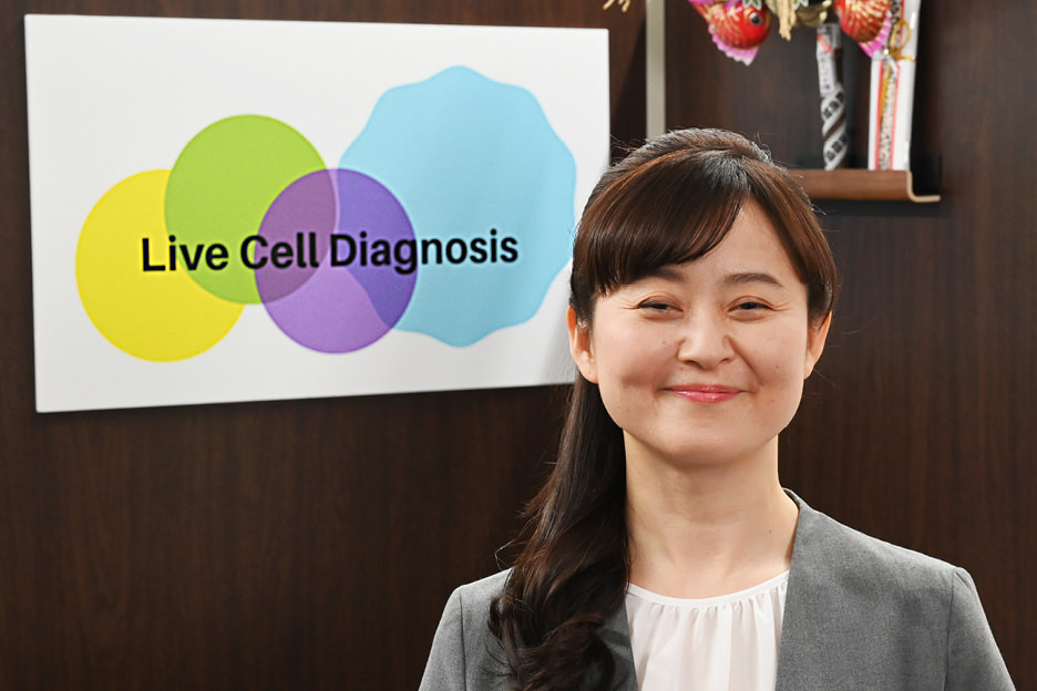
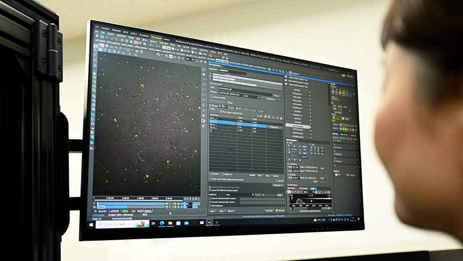
We recently spoke with Dr. Mai Yamagishi, who serves as the Representative Director of Live Cell Diagnosis Co., Ltd., a startup*1 company founded in 2019. The company focuses on a technology for imaging the secretion activity of living cells, known as Live-Cell Imaging of Secretion activity (LCI-S). "Our mission is to pioneer a novel approach to medicine by diagnosing live cells. At our company, we describe LCI-S as a technology that allows us to 'listen to the voices of cells'," explains Dr. Yamagishi. It is estimated that our bodies contain approximately 37 trillion cells, and they communicate with each other to maintain a healthy state. When one cell releases signaling molecules, various reactions occur as other cells receive these substances. "The types of signaling molecules that cells utilize to communicate with, as well as the timing and to which cells they communicate, are crucial for maintaining a healthy body," continues Dr. Yamagishi. "LCI-S allows us to visualize the signaling molecules released by cells in real-time, essentially enabling us to directly recognize the 'voices' of cells. This technology originated from the concept developed by the team of Professor Yoshie Harada at the Institute for Protein Research of Osaka University, and it has been realized with the support of many others."
Institute for Open and Transdisciplinary Research Initiatives, Osaka University
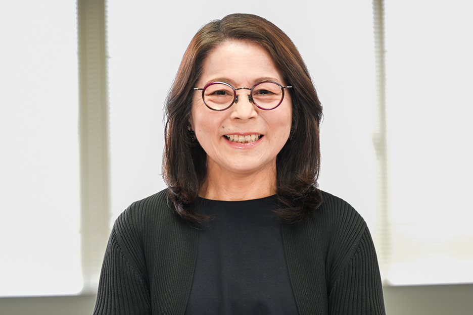
Prof. Yoshie Harada
*Position and affiliation mentioned are as of the time of the interview.
The research lab of Live Cell Diagnosis is located within a section of the Saitama Industrial Technology Center (SAITEC), which offers support for companies in technology, research and development, and business development. On the day of the interview, we also had the opportunity to speak with Professor Harada, who had come to the lab from Osaka. "I’ve known Dr. Yamagishi for around 20 years, first meeting her around 2002 when I was leading a clinical research lab at the Tokyo Metropolitan Institute of Medical Science," she recounts. Professor Harada elaborated, "In 1995, we developed single-molecule fluorescence imaging*2 technology using a fluorescence microscope equipped with total internal reflection*3 fluorescence to study the functions of individual protein molecules. Later on, we utilized this technology to conduct experiments to detect enzyme activity within individual cells and to identify antibodies secreted by cells. Dr. Yamagishi and her team continued these experiments and completed the current system," revealing the origins of LCI-S. Professor Harada also acts as an advisor to Live Cell Diagnosis.
- *1.A startup involves initiating a new business or project based on new technology or ideas, with a primary focus on solving societal issues.
- *2.Single-molecule fluorescence imaging is a technique that visualizes molecules using fluorescence, allowing observation of the molecular state within cells.
- *3.Total internal reflection refers to a phenomenon that occurs when light enters from a medium with a high refractive index into one with a lower refractive index. When light enters the boundary between the two mediums at an angle greater than a critical value, it results in all the light being reflected back into the first medium.
A microscope designed to meet the demands of emerging technologies
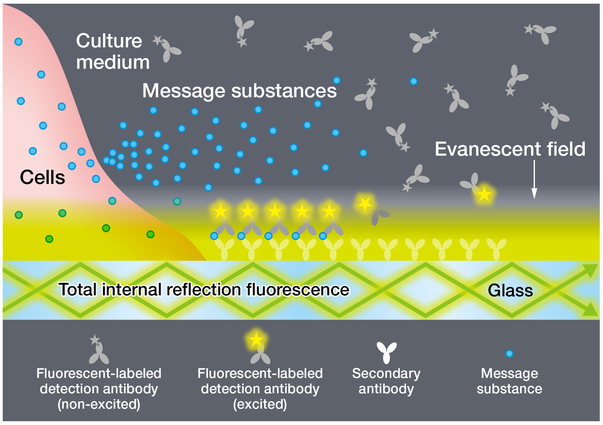
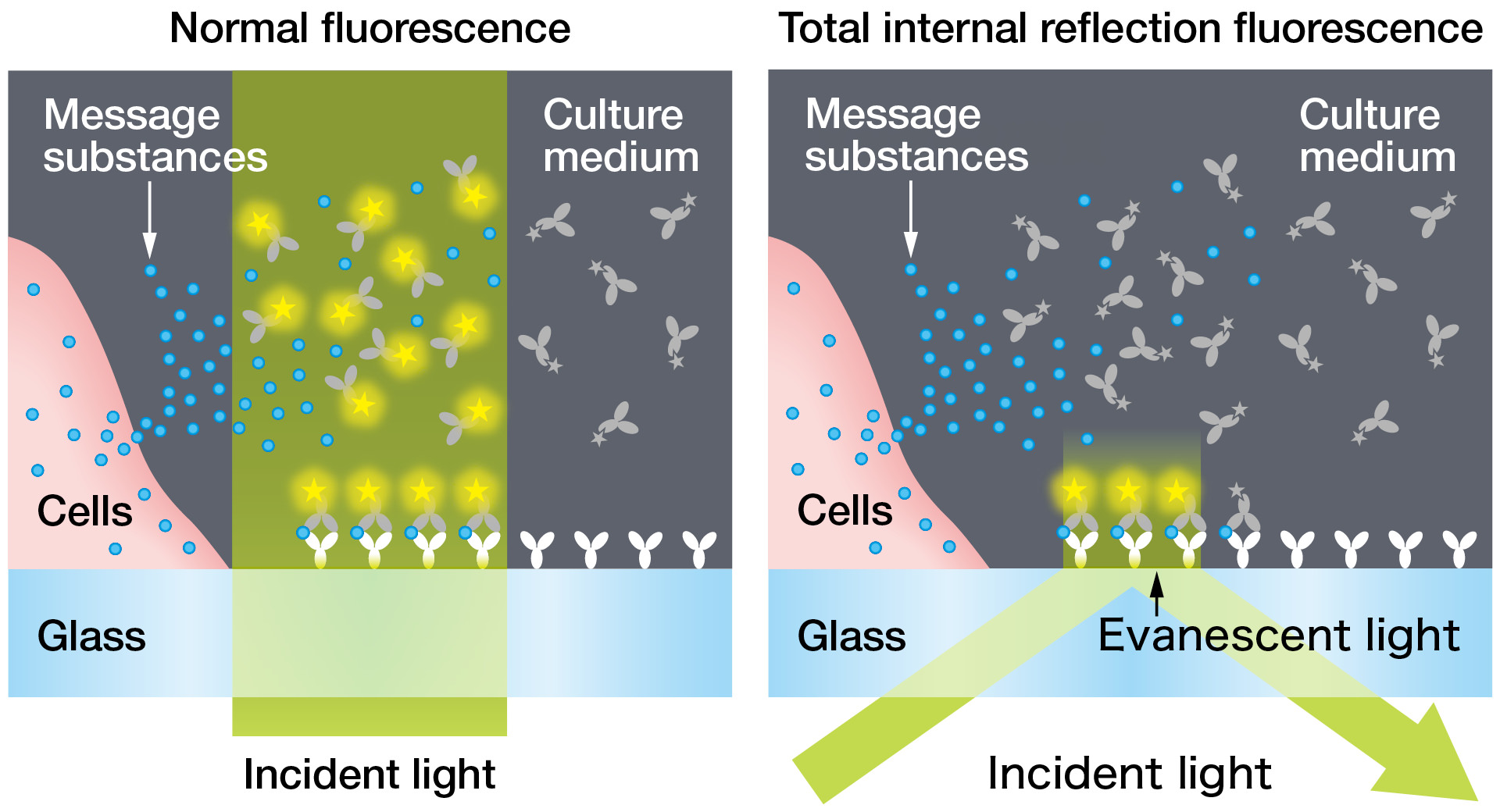
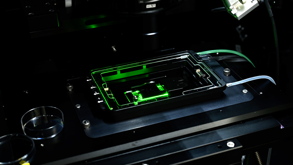
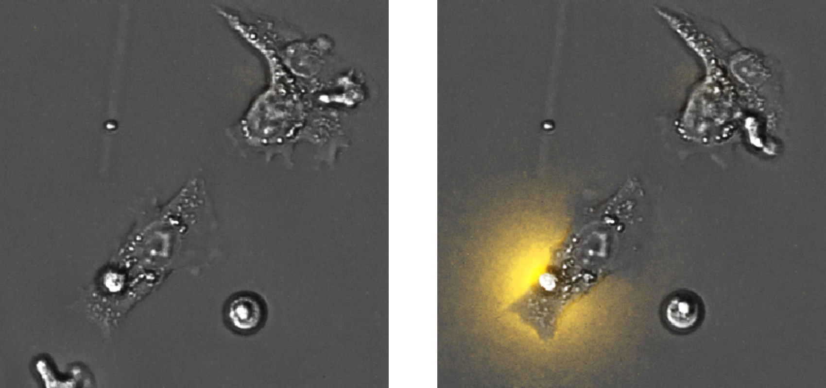
The principle behind LCI-S involves combining total internal reflection microscopy, commonly used in antigen-antibody testings*1 and single-molecule fluorescence imaging. Antigen-antibody tests utilize fluorescent dyes to visualize message substances released by cells on the surface of glass slides. However, to visualize these substances, excess fluorescent dyes are often added, which can complicate interpretation. This is because non-target substances can also emit fluorescence, necessitating the removal of the surplus dye in conventional practice. LCI-S employs total internal reflection fluorescence, directing light onto fluorescent dyes using a unique method. This approach ensures that only substances near the glass surface fluoresce, thanks to the phenomenon of evanescent*2 light, enabling clearer detection without the need to remove excess dye.
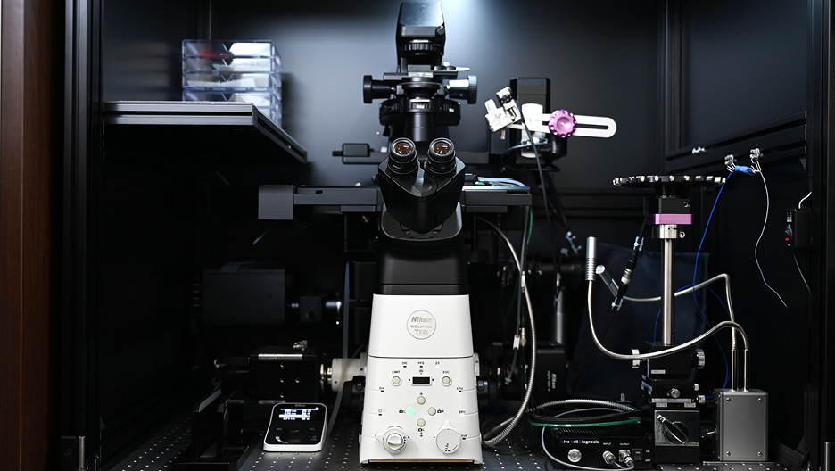
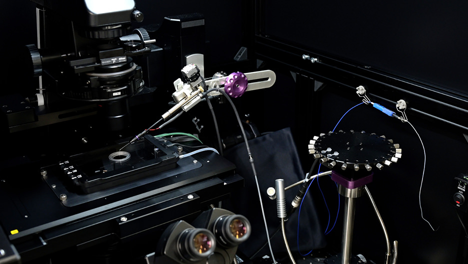
"Currently, at Live Cell Diagnosis, we rely on Nikon's inverted microscope, the ECLIPSE Ti2-E, which allows us to observe living cells samples as they are, as the foundation for all our research and development," explains Dr. Yamagishi. Being electrically operated, it can automatically switch settings and control the stage, greatly empowering observations through capturing images of multiple samples and conducting time-lapse observations. "Initially, we used a different microscope, but around 2009, when we tried Nikon's microscope, I remember being amazed at how fast we could acquire images. We’re also very pleased with the Nikon microscope’s features, such as its wide field capability. And besides its great features, the stable performance of its objective lenses and quality, the dedicated support from Nikon's representatives, who attentively addressed our concerns and needs, was a significant factor in our decision to adopt the current microscope," she adds with appreciation. Interestingly, the same microscope system has been implemented in Professor Harada's lab in Osaka. Sharing the same system facilitates easy understanding of research and development directions and enables quicker and more accurate responses when issues arise.
- *1An antigen-antibody test is a method for detecting and measuring small amounts of substances by utilizing the specific and strong bonds between antigens and antibodies.
- *2Evanescent light is a unique type of light that penetrates into the lower refractive index material under conditions of total internal reflection.
For the well-being and happiness of everyone
Lastly, we asked both about their future prospects. Dr. Yamagishi shared, "Currently, we're conducting foundational research to showcase specific examples of how LCI-S can contribute to healthcare, and we're also progressing with equipment development to enhance the usability of LCI-S. For instance, our goal is to finalize a diagnostic device capable of promptly diagnosing cells collected from patients directly in medical settings. Additionally, given LCI-S's ability to diagnose individual patients' cells, we envision it aiding in the realization of precision medicine tailored to each individual's condition." She also revealed their plans to establish a platform enabling analysis of data obtained from tests on clouds, and provide globally recognized evaluation criteria based on LCI-S in the future.
Professor Harada further commented, "With this technology, we can visualize the natural process of cell secretion, such as the abundant secretion of cytokines that trigger inflammation or allergies from activated immune cells. Moreover, LCI-S has the potential to contribute to areas like drug discovery and regenerative medicine, as it can be applied to cells obtained from mice, human clinical samples, ES cells, iPS cells, and more. Going forward, we intend to continue conducting experiments that demonstrate the effectiveness of LCI-S and work towards making this technology more widely accessible for utilization.
Dr. Yamagishi and her team are working on initiatives aimed at bringing new schemes to healthcare and drug development through innovative technologies, contributing to people's health and well-being. Nikon will continue supporting their efforts via providing a variety of microscopy technologies, as well as through our personnel and services, to ensure their ongoing success.