Accurate measurement MSC cell number and growth rate in an non-exfoliating and fast way
Point
Changes in the number of human somatic stem cells , such as MSCs (mesenchymal stem cells ), can be accurately measured over time without having to remove cells from culture.
Overview
Human somatic stem cells, such as MSCs , can only undergo a limited number of cell-division cycles, and the proliferation rate declines following each passage. Therefore, it is important to have a clear understanding of the number of passages appropriate for the cell type in question during process development. If improper culture conditions change cell characteristics, anomalies in proliferation may result. The cell proliferation rate is thus an important index for the evaluation of culture conditions.
Sometimes, proliferation cannot be confirmed in primary culture. In such a case, the cell count must be monitored over time to determine whether or not to continue that culture. Operator judgment is generally informed by visual observation, which may compromise sensitivity.
Current
Issue-1
Traditional cell counting methods require a lot of work and damages cells.
Traditional cell counting methods, for example using a hemocytometer, require dissociation of cells from the culture vessel. This operation requires significant time and energy on the part of the operator, and also damages cells.
Issue-2
It is impossible to continuously count the number of cells.
Ideally, cell proliferation rate is measured over a specified time period at regular intervals, and without removing cells from the vessel. Measurement method requiring cellular trypsinization is invasive, and require many samples seeded under equivalent conditions. Cells are counted by invasive treatment with every measurement, posing a challenge to collecting a sufficient number of samples.
Solution
Non-invasive phase contrast image analysis allows for the number of cells in culture to be measured over an extended period. Cells may be moved to the next step of the culture process without being wasted.Cell manufacturing processes for regenerative medical products often require more cells than can be readily secured. Therefore, a non-invasive, non-staining inspection is desired in order to conserve cells.
In autologous cell culture, it is difficult to implement a strict manufacturing process, so a procedure that is flexible within a predefined allowance should be designed in order to be able to adapt to the observed state of proliferation.
For such process control, non-invasive measurement using automatic image acquisition and analysis enables real-time monitoring of cell proliferation condition.
In developing a flexible manufacturing process, a system such as BioStudio-T*, a cell observation system which may be installed inside an incubator, reduces cell stress while enabling real-time observation. The entire vessel can be monitored using the image stitching function of these systems.
*This product has been discontinued. If you have any questions about cell cultures, assays, or products, please contact us using the contact form.Cell image analysis software Cell Analysis Module
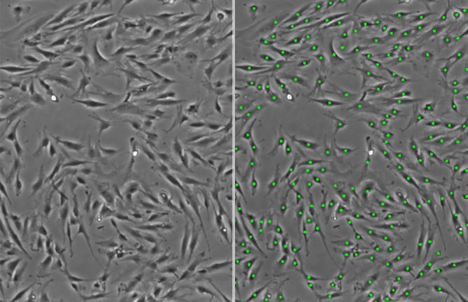
Measurement of MSC number by non-invasive phase contrast image analysis (human bone marrow-derived MSC UE7T-13 (JCRB1154))
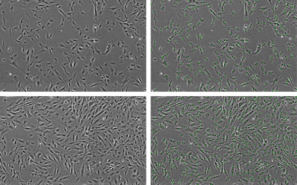
Cells even at different cell densities can be detected in images.
Left: phase contrast image, right: MSCs detected through image analysis
Top: low density, bottom: mid density
Utilization scene
As an evaluation criterium for establishing cell culture processes and procedures
- Validation of culture conditions
- Understanding the passage number and population doubling number, medium composition, passaging and procedures, etc.
- Detection of cells which proliferate abnormally due to transformation
- Comparison of proliferation among cell lines
Cell image analysis software
NIS-Elements Cell Analysis Module
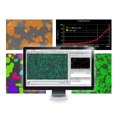
The following application softwares are for the purpose that specific image analysis can be performed simply using Nikon's Image Analysis Software in the field of research and development. As we have developed image analysis algorithms for many years and accumulated know-how, we have started to provide add-on Modules.
Click here for detailsNikon will contribute to solving your cell culture issues with its image analysis
techniques and know-how on cell quality evaluation.
Click hereInquiry Form
Share this page