Elucidating the brain mechanisms that control perception, cognition and decision-making, is one of physiology and medicine’s major challenges. In spring 2021, a joint research group at the Laboratory for Haptic Perception and Cognitive Physiology at RIKEN Center for Brain Science developed a microscope that captures large-scale activities with a wide field-of-view, and high-resolution, high-speed imaging. Nikon’s microscope technology contributed to this groundbreaking project, in which laboratories, universities and businesses worked together.
A microscope that captures a ‘large-scale network’ of the brain
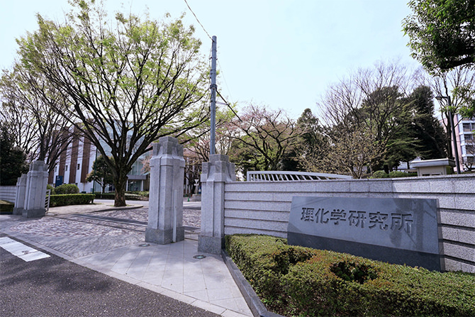
The RIKEN Center for Brain Science is Japan’s largest comprehensive research institution specializing in high-quality research over a diverse range of scientific disciplines such as physics, engineering, chemistry, biology and medical science. We visited its base facility in Wako city, Saitama Prefecture, Japan.
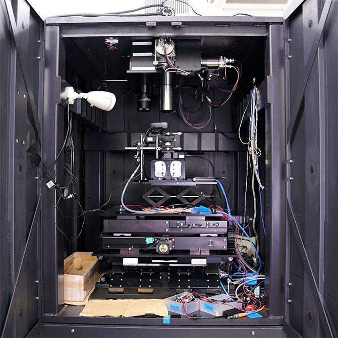
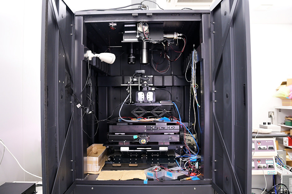
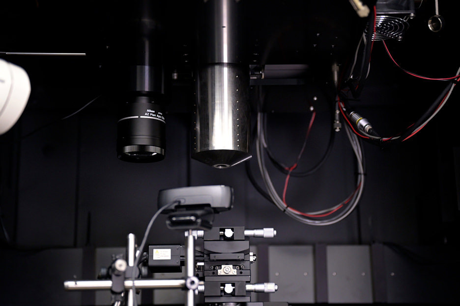
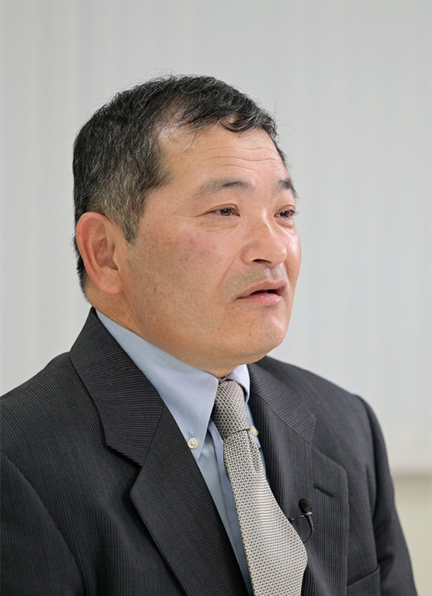
Masanori Murayama, PhD, team leader of RIKEN’s Laboratory for Haptic Perception and Cognitive Physiology spoke with us. A joint research group led by Dr. Murayama developed the FASHIO-2PM special laser microscope*1. The FASHIO-2PM is an epoch-making microscope that can capture the activity of more than 16,000 nerve cells in the cerebral cortex of mice with a wide field-of-view 36 times that of conventional microscopes, and which has also succeeded in capturing moving images. This extremely wide field-of-view, the large number of recorded cells, and a shooting speed that realizes recording of moving images make it the world’s largest and fastest*2 laser microscope — an achievement that has attracted the attention of many researchers.

Laboratory for Haptic Perception and Cognitive Physiology
Team Leader
Masanori Murayama, PhD

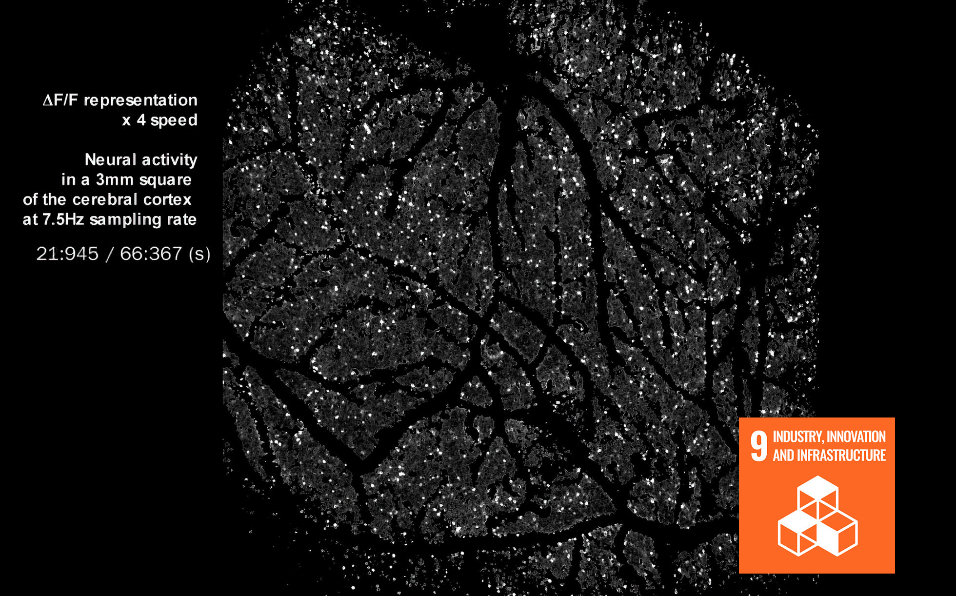
So, what kind of new vision has the FASHIO-2PM brought to elucidating brain functions? The video below shows the neuronal activities of a mouse brain. The shining dots that look like stars in the night sky are neurons expressing fluorescent proteins called calcium sensors, generated in the mouse’s body. When subjected to excitation*3 by the laser light of the FASHIO-2PM, they emit light. The sparkle becomes more pronounced as the activity of nerve cells becomes greater, and you can clearly see the real state of brain activity.
Movie courtesy: RIKEN Center for Brain Science.
Although it has been possible to monitor brain activity with a conventional laser microscope, it was limited to a very narrow area. To overcome this issue, wide-field microscopes have been developed recently. However, due to the trade-off relationship between resolution and sensitivity, the images from these microscopes tended to be dark and unclear, with a shooting speed of about one frame per second, making it impossible to accurately track instantaneous changes in brain activity. The FASHIO-2PM can capture a high-sensitivity image of a wide 3x3mm field-of-view with cell-level resolution that shows multiple areas of the brain simultaneously. It can also capture instantaneous changes at a speed of 7.5 images per second. “When I played this video at a symposium, people were amazed. So, I feel that this microscope is really something special,” explained Dr. Murayama during the development announcement.
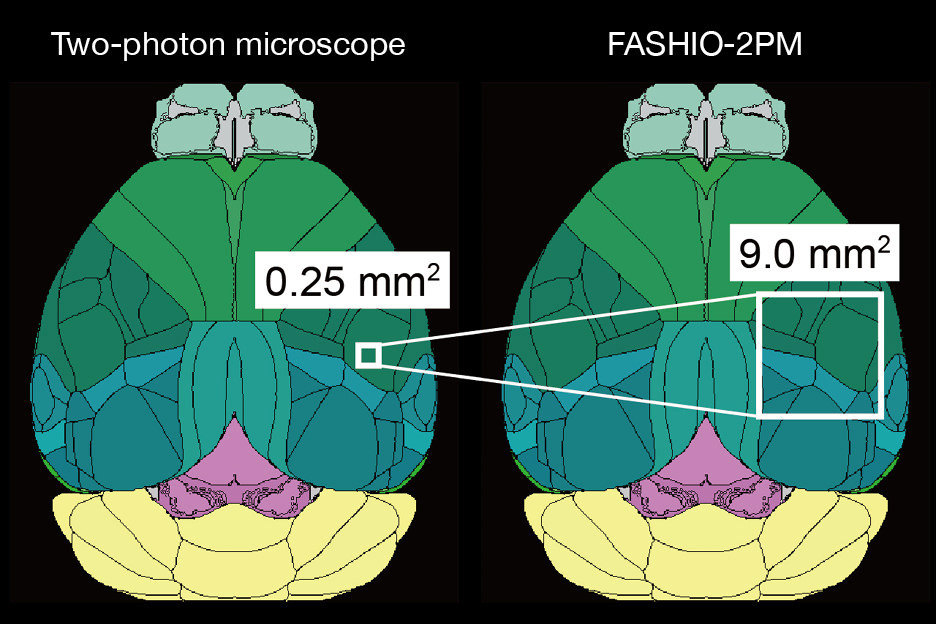
Image courtesy: RIKEN Center for Brain Science.
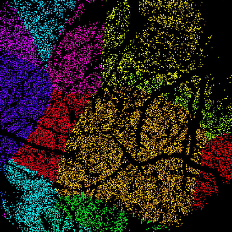
Image courtesy: RIKEN Center for Brain Science.
- *1A laser microscope is an optical microscope that uses laser light as a light source for observation. Normally, LEDs and halogen lights are utilized. The FASHIO-2PM employs two kinds of laser beams.
- *2Source: RIKEN press release on April 20, 2021. (Japanese only)
www.riken.jp/press/2021/20210420_1/![[Open in a new window]](/common/images/icon/icon_blank.svg)
- *3Excitation refers to the condition of an atom or molecule when it absorbs energy such as heat or light and moves from its original state to a high-energy state.
Cutting-edge technology and a solid partnership
The joint research group consisted of two research institutes, five universities, and three companies*. Nikon also participated and contributed to the development of the FASHIO-2PM. We also talked to two Nikon engineers who were involved in the design process. Because this microscope can perform ultra-high-speed scanning while irradiating a pulsed laser that blinks repeatedly at very short intervals, it enables the capture of images by controlling a special mirror scanner that reflects light at high speed and with high accuracy. That is why chief engineer Yoshio Kuroiwa was needed for the designing of the electrical hardware. “We carefully listened to Dr. Murayama’s required specifications and requests, and repeated discussions on methods for realization, equipment configuration, and roles of team members”, says Mr. Kuroiwa, who has been involved in the development of laser microscopes for more than 30 years. In reference to chief researcher Masaru Horikoshi, who is in charge of firmware design, he continues, “It’s all software that makes electrical hardware function, and we’ve always worked closely with Mr. Horikoshi.”

Technology Solutions Sector
Designing Department
2nd Designing Section
Chief Engineer
Yoshinori Kuroiwa
Mr. Horikoshi designed the firmware inside the controller, the software for setup, and the specifications of the application software to be used by researchers. When asked about the joint research group that brought a variety of cutting-edge technologies and knowledge together, Mr. Horikoshi explains, “Everyone was overjoyed when an image was taken for the first time. It was a united team that tackled the challenge of development while sharing such excitement.” He continues, “Regarding the software used by researchers, we carefully listened to what Dr. Murayama needed, and created something that is fairly easy to use.”
When asked about the factors that enabled them to meet the specifications required by Dr. Murayama, they explain that they were able to clear such high demands because of the microscope development technology and know-how that Nikon had accumulated over the years. The same is true for other team members. It could be said that the FASHIO-2PM was born from a combination of both the advanced technological knowledge of each staff member and a sense of unity.
- *Two laboratories at RIKEN and the National Institute for Physiological Science. Five universities: Tokyo University, Tokyo Polytechnic University, Juntendo University, Tohoku University, and Kyushu University of Health and Welfare. Three businesses: Fob Co., Ltd., Hamamatsu Photonics K.K., and Nikon Co., Ltd.
Technology Solutions Sector
Designing Department
3rd Designing Section
Chief Engineer
Masaru Horikoshi
A new challenge extended from a new perspective
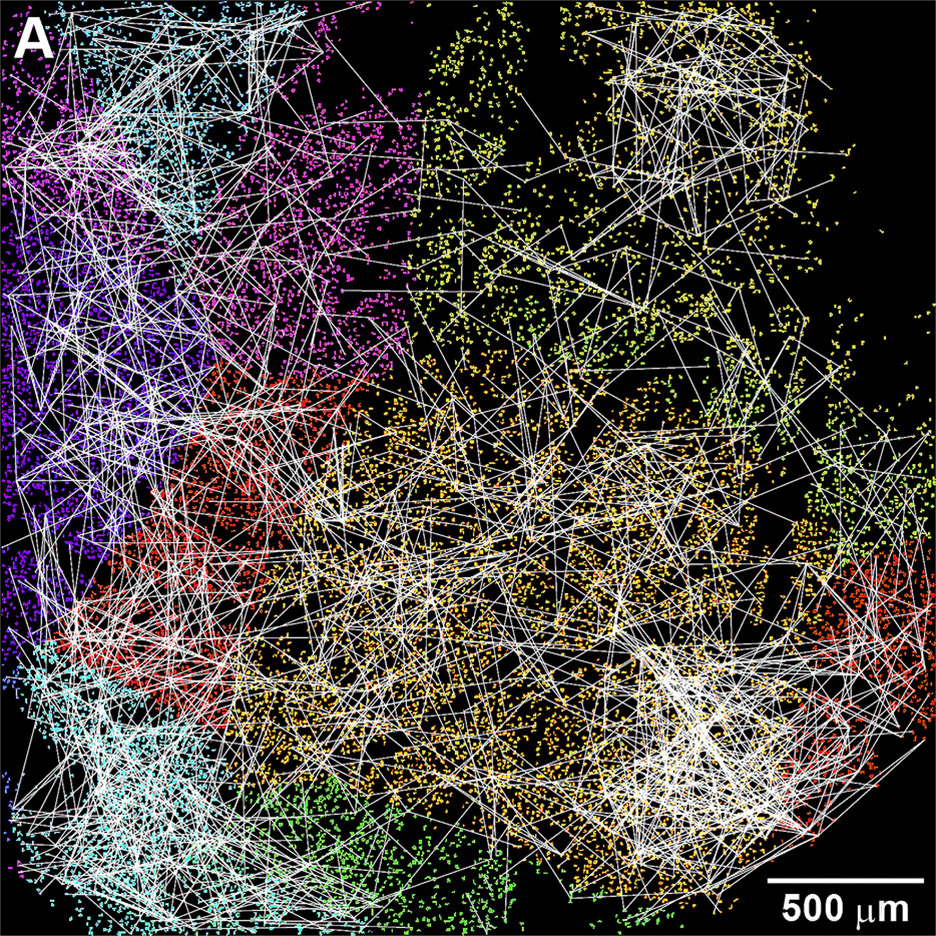
Functional coupling forms clusters within the same brain field.
Image courtesy: RIKEN Center for Brain Science.
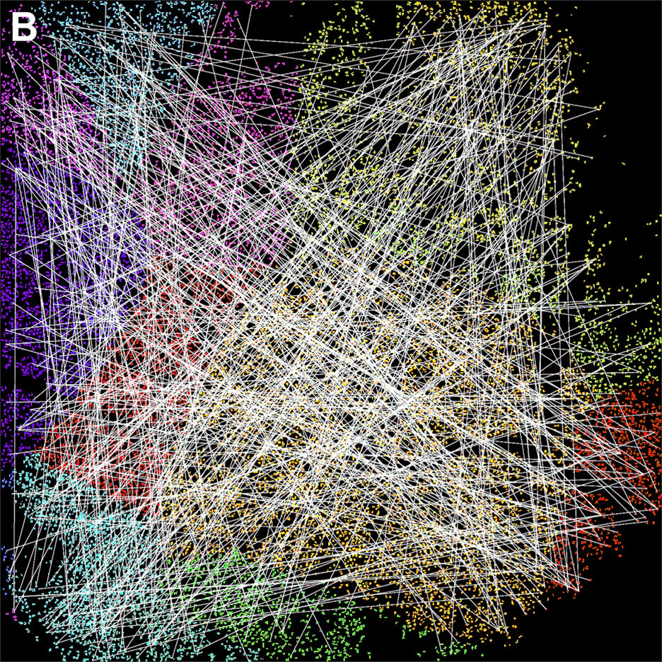
Functional coupling forms clusters across the brain field.
Image courtesy: RIKEN Center for Brain Science.
Dr. Murayama and his team have further advanced research and visualized the nerve cells and functional coupling of the brain. These are shown in the two images above which also reveal that the neocortex has the very efficient information-processing property of not only being able to coordinate at short distances, but also with cells in distant locations. They also found hub-like neurons that cooperatively work together with more than 100 neurons. These discoveries have drawn worldwide attention and brought a new perspective to the field of brain science.
“Tactile perception is a basic function of the brain,” explains Dr. Murayama, “but even the mechanism behind it isn’t yet clear. If we can understand this, we will be able to clarify the mechanisms of decision-making, movement, and execution. Elucidating these functions is the ongoing goal of our work.”
As these efforts continue, Nikon’s technology will also be there, contributing to the challenge of uncovering the secrets of the brain.