The inner workings of our brains are a subject still shrouded in a great deal of mystery. However, it is known that the human brain contains a vast number of nerve cells, linked by the synapses between them.
By studying the proteins inside synapses within the brain, it was discovered that there is an immense variety of synapses. Nikon’s microscope technology is used for observing the proteins of synapses as broadly — and in as much detail — as possible.
Coloring synapses to create a map
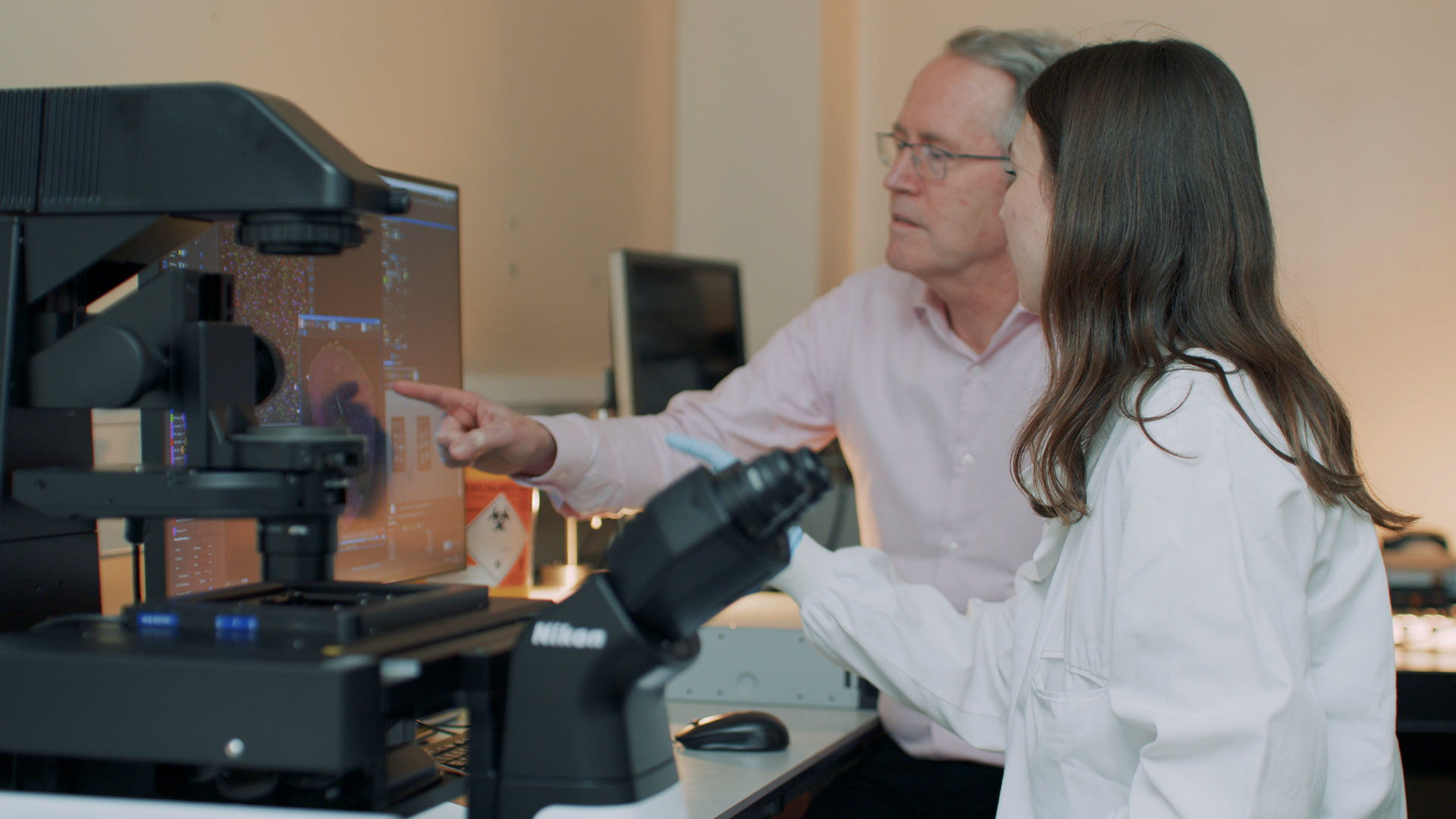
For over 30 years, Professor Seth Grant at the University of Edinburgh has been studying the molecules inside synapses to help determine the relationship between gene mutations and numerous kinds of brain disease.
“The brain is the most complex object in the universe, and understanding how it works is one of the greatest frontiers in science today. That’s because we need to understand how thinking works, and then how the brain goes wrong and causes hundreds of different brain diseases. So, understanding the basic science of the brain is an absolutely key area of modern science,” Professor Grant explains.
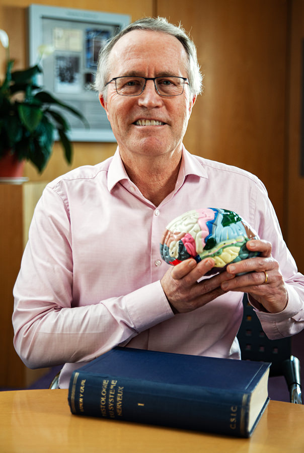
Centre for Clinical Brain Sciences
Professor of Molecular Neuroscience
Seth G.N. Grant FMedSci FRSE
*Job title and responsibilities at time of interview.

right) Microscope image of mouse brain
By focusing on understanding the normal functions of the brain, Professor Grant uses his research to further understand the various kinds of brain diseases affecting millions of people worldwide. By undertaking a tremendous amount of work analyzing the proteins within the billions of synapses in the human brain, his team discovered that each activity and experience stimulates certain synapses which then corresponds to different electrical responses. Utilizing this crucial data, his team has employed artificial colorization for each individual protein to create maps of the brain in mice. “If we map [the proteins], we can determine where diseases occur in the brain,” he explains. Through this research, the team has revealed the biological basis of many genetic disorders affecting the brain, including schizophrenia, characterized by the mutations in the synapses.
Professor Grant continues, “I think most people will be very interested to understand how brain disorders work and to have new therapies for them. So, we envision that one area of our work is that we will discover which synapses in the brain are damaged in each of the different diseases. And that's going to be very important going forward because there can be new therapies and new drugs based on those kinds of discoveries.”
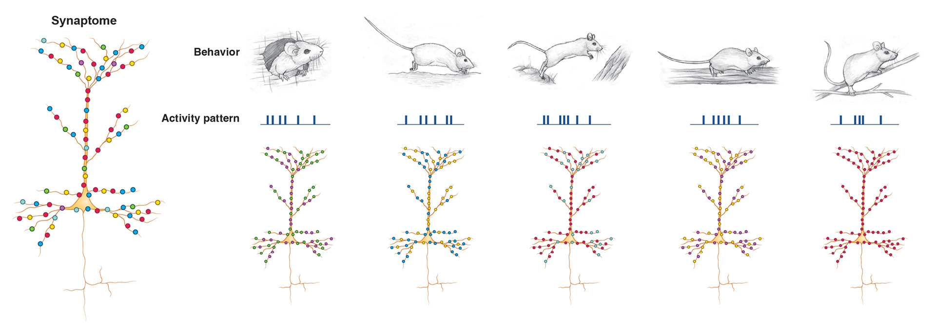
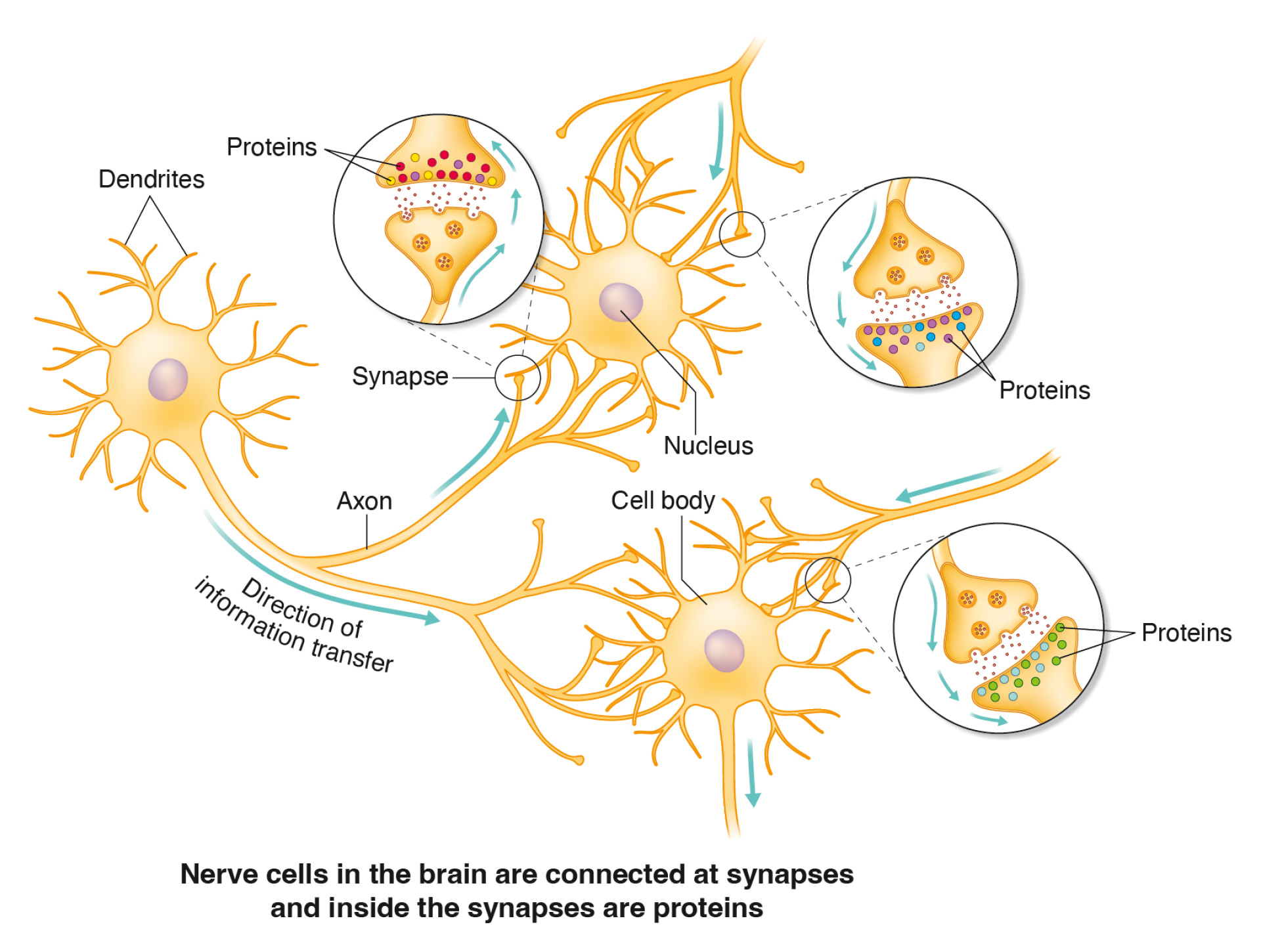
Nerve cells in the brain have a dendritic tree that is covered with many synapses, and these synapses possess molecular differences (shown by the colored spots in the above diagram). As animals behave and move through the world there are patterns of electrical activity in their brains that activate the different synapses (shown by the different colors of the synapses beneath each behavior and activity pattern).

Technology to broadly and effectively observe synapses
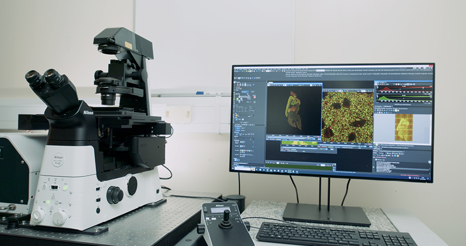
Unraveling such molecular mechanisms can shed light on how various external factors influence our brains and, consequently, our behavior. In order to achieve such a monumental task, it requires analyzing in detail the extremely complicated human brain – examining trillions of synapses as broadly as possible. “The objective of our research is to look through the microscope at each of these individual synapses, which are just a fraction of the width of a human hair. But we don't want to just observe one or two of them – we want to see billions of them in the brain. And we need a specialized kind of microscope for that purpose, which is capable of visualizing individual synapses and the proteins in them,” Professor Grant explains. That’s where Nikon’s microscopes entered the picture to aid Professor Grant in his research.
“This Nikon Ti2-E with CSU-W1 enables us to capture images of brain tissue at both very high magnifications and an expansive scale simultaneously. With this microscope, we can effectively capture images of billions of synapses, allowing for the quantification of proteins within them and detailed analyses of their size, shape and other features,” he explains.
Professor Grant continues, “As a result, it provided us with a groundbreaking opportunity to analyze, for the first time, the diversity of various synapse types and their spatial distribution in brain tissue samples.” The valuable data provided by this incredible technical capability realized the creation of the aforementioned intricate brain maps.
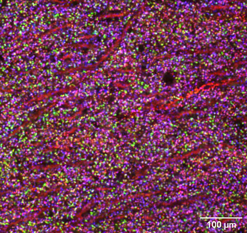
Image of mouse brain tissue showing individual synapses as colored puncta. Three different proteins were labelled with green, red and blue dyes. The diversity of synapse types shown by the rainbow of colors results from the combined expression of these three proteins.
Opening up new possibilities for brain disease treatment
Professor Grant’s efforts have helped support the refinement of new technologies that facilitate the study of human brain pathology at the synaptic level. He is now convinced that his approach holds promise for further application across a myriad of brain diseases. “Through this method, we have unveiled previously unseen pathologies in the brain associated with these disorders,” he says. “We are optimistic that these newfound insights will guide us towards novel therapeutic strategies and innovative methods for monitoring the initiation and progression of these diseases in clinical settings.”
In closing, Professor Grant shared his expectations for Nikon in the future – specifically in terms of developing progressively faster spinning-disc confocal microscopes capable of swiftly scanning larger areas of tissue. “This capability will enable a microscopic-level understanding of the intricate complexity of the human brain, and I think that will be an important area for innovation,” he concludes.
We at Nikon will continue to make determined efforts to meet Professor Grant’s expectations and strive for future progress in the vital field of brain research.