
Image Analysis – useful techniques for improving cell culture processes
Regenerative medicine continues to mature and gain in popularity. Unlike traditional medicines, the cells themselves act as therapeutic active substances.
This poses new process control challenges for ensuring the reliable culture of cells, the accurate evaluation of their quality, and for obtaining target levels of satisfactory cells without extensive purification processing.
For that reason, it is important to fully understand the characteristics of the cells, and to evaluate quality both during the manufacturing process and in the final product using indicators that are appropriate for the specific cell types being cultured.
Furthermore, it is also necessary to construct a highly repeatable manufacturing process utilizing these indicators.
Therefore, this article describes Image analysis techniques which have been recently introduced for the development of cell manufacturing processes and quality control.
History of Regenerative Medicine and Its Future Role

Regenerative medicine is a medical field that uses stem cells derived from either the patients themselves or healthy donors in order to replace, repair, or regenerate damaged cells, tissues, or organs as part of the treatment or prevention of various diseases.
Regenerative medicine is attracting attention because it may be able to treat previously intractable diseases and injuries, such as spinal cord injury, severe heart failure, and Parkinson's disease, which are difficult to treat with conventional surgical methods and drugs. Additionally, the number of patients that develop age-related diseases, such as age-related macular degeneration, is expected to grow in our aging society, resulting in a more pressing need for effective treatments. Regenerative medicine is expected to establish its own genre of medical treatment, and its applicability to grow as regenerative medical products continue to be developed with demonstrated safety and efficacy for a wide variety of diseases and injuries.
Regenerative medical products have been actively developed in the world for over 30 years. The raw materials have mainly been somatic stem cells and MSCs (mesenchymal stem cells), but since the establishment of human ES (embryonic stem) cells in 1998 and human iPS (induced pluripotent stem) cells in 2007, various types of cells are now being utilized for different use cases.
In 2007, the EU classified regenerative medicine products as belonging to the category of Advanced therapy medicinal products (ATMPs), which is within the category of conventional pharmaceutical products (Regulation (EC) No 1394/2007), and regulations have been established. In the United States, regenerative medicine products are mainly regulated as products that fall into the category of Human Cells, Tissues, and Cellular and Tissue-derived Products (HCT/Ps) within the category of Biologics or medical devices (21CFR1271, 351HCT/P), and regulations are in place. In 2014, the Pharmaceuticals, Medical Devices, and Other Therapeutic Products Act (PMD Act) and the Act on the Safety of Regenerative Medicine (RM Act), which are both specifically for regenerative medicine, were enacted.
Thanks to the expansion in cell options and legislation to help facilitate the development and distribution of regenerative medical products described above, startups and major pharmaceutical companies are entering into the market (with peripheral businesses growing in step), establishing a foundation upon which the development of regenerative medical products is set to grow significantly.
High Quality Control Standards Are Required for Cell-Based Regenerative Medicines
In regenerative medicine, cells are transplanted into a patient, and treatment is performed by the action of the cells. In other words, the cells themselves become drugs. Just as conventional drug manufacturing is under strict control in accordance with regulations, strict control is required for the manufacturing of cultured cells for therapeutic purposes.
To be approved by the regulatory authorities to market regenerative medical products, a manufacturing system that conforms to (current) Good Manufacturing Practice ((c)GMP) and (current) Good Tissue Practice ((c)GTP), or Good Cell and Tissue Practice (GCTP) must be developed and sufficient data provided on the efficacy and safety of the products. (c)GMP and GCTP are quality control standards established by the each country/region’s pharmaceutical agencies such as FDA, EMA, MHLW in order to ensure the quality of regenerative medical products. Compliance requires the establishment of SOPs (Standard Operating Procedures), in addition to the recruitment, education, and training of human resources.
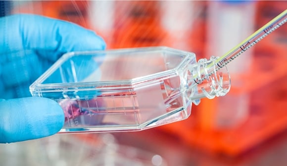
Real World Issues in Cell Culture
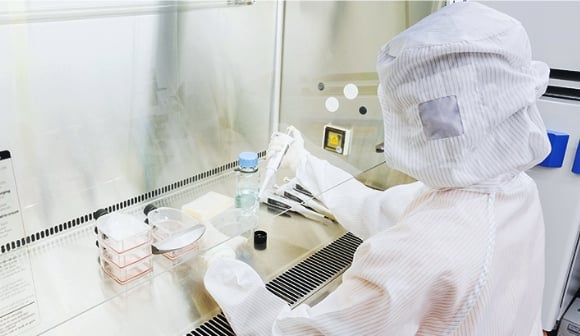
In many cases, the current manufacturing processes of regenerative medical products are manually performed by human operators. Manual work has its own advantages, especially in the development phase, where flexibility is often required. However, there are also certain issues that arise due to the dependence on an individual's skills.
Each operation of the cell culture process, such as cell suspension, seeding, medium change, and enzymatic treatment, varies with different culture systems and is dependent on the operator in terms of the time required and the force applied in each operation.
These factors result in differences in the physical impact experienced by the cells, which can lead to variability in their properties. Many cell properties can be assessed using indicators such as various morphological, quantitative and behavioral characteristics, including the number of cells in culture. Furthermore, indicators based on such characteristics are effective in many situations during the in-process control to improve for improving the stability and consistency of production. Examples include the degree of cell proliferation and cell differentiation.
Currently, these indicators are visually determined by operators using a microscope. Measurement of cell state or other characteristics by a phase contrast microscope, using cell morphology as an indicator, is a non-destructive inspection method for measurement and analysis that doesn't waste precious cells. It is particularly beneficial in the development and manufacturing of regenerative medical products, which often involve small numbers of cells. Additionally, in the manufacturing process of regenerative medical products, the decision to proceed to the next step is quite often made based on cell state. If a measurement method requires time to determine the cell state, the production process has to stop until the measurement result becomes available, a period during which cells may become damaged. Even if the culture is continued without stopping the process, there is a risk of failing to make an accurate judgment because a difference between the state of the cells at the time of sampling already differs from the current state. Therefore, it is crucial to have a measurement method that uses cell morphology as an indicator in order to measure quickly.
Differences in technical skills among operators can affect cell health, as well as influence critical decisions that must be made during the culture process in order to avoid inconsistencies that may lead to compromised cell quality. Even for a single operator, we cannot deny the risk of variability in procedures and judgments. These risks are only exacerbated by the current physical and mental condition of the operator, with even further variability introduced by their surrounding work environment. In order to prevent these differences and risks, it is critical to properly secure and educate these operators, which requires significant time and cost. However, in reality, these factors may never be fully controlled for.
Quantification and automation of cell quality assessment with image analysis technology
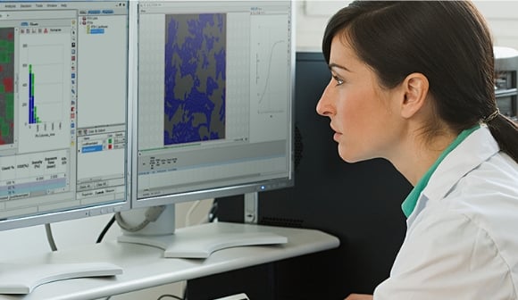
Where should we start if we want to culture cells that meet target quality standards in a more reliable manner than we currently have? As a first step, how about quantifying as much as possible important indicators used in the processing and quality control of cell cultures that are currently dependent on the operator? Once quantitative standards are established, cell states and the validity of the manufacturing process can be objectively evaluated. Improvements to the manufacturing process should result in more reliable and consistent cell quality. Quantitative criteria are best for process automation and will help lead to partial automation of the cell quality evaluation process that previously relied on humans.
Methods for evaluating the characteristics of cultured cells from cell images can be considered as consisting of combinations of three techniques: "photography," "recognition," and "evaluation." To automate the "recognition" and "evaluation" processes, information on the morphological and behavioral characteristics of the cells needs to be analyzed using image analysis software for conversion into quantitative numerical values. If important unbiased indicators related to the cell manufacturing process can be identified using such information, then we will come closer to realizing both quantitative cell quality control and manufacturing process control.
This website introduces problems that are likely to occur in the field and "improvement examples" of solutions to those problems that use image analysis.
Contribution of image analysis to automation of the cell culture process

In order to evaluate whether cells and their manufacturing processes meet the appropriate quality standards required for regenerative medicine products, it is very important to introduce image analysis technology for in-process quality control and quantification of evaluation criteria. Quantification activities will also be very useful when automating the manufacturing process.
There is an increasing need for mechanization and automation of some or all of the manufacturing process in order to achieve reliable production of cells of consistent quality. Such efforts have already begun. Mechanization and automation could potentially solve many challenges by improving the quality of cultured cells, reducing the amount of work done on site, reducing cost, and avoiding the risk of bacterial contamination thanks to the absence of human intervention. However, when replacing manual culture processes with mechanization and automation, the challenge becomes making sure that the process is equivalent to that performed by humans.
What becomes important is the accumulation of data at the research and development stages. If quantitative information regarding the morphological and behavioral characteristics of cells is collected during the culture process, it will be extremely useful in assessing the effects of changes in the process.
In the development of regenerative medical products, whose market is expected to expand rapidly, it is fair to say that the use of image analysis technology is the key to achieving a competitive edge in the global marketplace and enhancing business competitiveness.